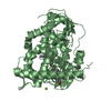
 Movie
Movie Controller


+ Open data

- Basic information
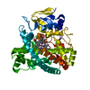
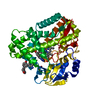
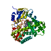
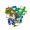
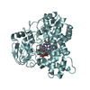
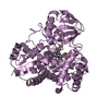
| Entry | Database: PDB / ID: 5l1p | ||||||
|---|---|---|---|---|---|---|---|
| Title | X-ray Structure of Cytochrome P450 PntM with Pentalenolactone | ||||||
 Components Components | Pentalenolactone synthase | ||||||
 Keywords Keywords | OXIDOREDUCTASE / PntM / cytochrome P450 / pentalenolactone | ||||||
| Function / homology |  Function and homology information Function and homology informationpentalenolactone synthase / pentalenolactone biosynthetic process / oxidoreductase activity, acting on the CH-CH group of donors, iron-sulfur protein as acceptor / oxidoreductase activity, acting on paired donors, with incorporation or reduction of molecular oxygen / antibiotic biosynthetic process / monooxygenase activity / iron ion binding / heme binding Similarity search - Function | ||||||
| Biological species |  Streptomyces arenae (bacteria) Streptomyces arenae (bacteria) | ||||||
| Method |  X-RAY DIFFRACTION / MOLECULAR REPLACEMENT / Resolution: 2.28 Å X-RAY DIFFRACTION / MOLECULAR REPLACEMENT / Resolution: 2.28 Å | ||||||
 Authors Authors | Duan, L. / Jogl, G. / Cane, D.E. | ||||||
 Citation Citation | Journal: J.Am.Chem.Soc. / Year: 2016 Title: The Cytochrome P450-Catalyzed Oxidative Rearrangement in the Final Step of Pentalenolactone Biosynthesis: Substrate Structure Determines Mechanism. Authors: Duan, L. / Jogl, G. / Cane, D.E. | ||||||
| History |
|
- Structure visualization
| Structure viewer | Molecule: MolmilJmol/JSmol |
|---|
- Downloads & links
-Download
| PDBx/mmCIF format | 5l1p.cif.gz | 184.9 KB | Display | PDBx/mmCIF format |
|---|---|---|---|---|
| PDB format | pdb5l1p.ent.gz | 144.5 KB | Display | PDB format |
| PDBx/mmJSON format | 5l1p.json.gz | Tree view | PDBx/mmJSON format | |
| Others |  Other downloads Other downloads |
-Validation report
| Arichive directory | https://data.pdbj.org/pub/pdb/validation_reports/l1/5l1pftp://data.pdbj.org/pub/pdb/validation_reports/l1/5l1p | HTTPS FTP |
|---|
-Related structure data
| Related structure data |  5l1oC  5l1qC  5l1rC  5l1sC  5l1tC 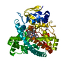 5l1uC  5l1vC  5l1wC  2x9pS C: citing same article ( S: Starting model for refinement |
|---|---|
| Similar structure data |


-Links
 PDBj
PDBj
- Assembly
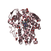
| Deposited unit | 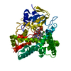
| ||||||||
|---|---|---|---|---|---|---|---|---|---|
| 1 |
| ||||||||
| Unit cell |
| ||||||||
| Components on special symmetry positions |
|
-Components
| #1: Protein | Mass: 44504.809 Da / Num. of mol.: 1 Source method: isolated from a genetically manipulated source Source: (gene. exp.) Streptomyces arenae (bacteria) / Strain: Tu469 / Gene: pntM / Plasmid: pET28a / Production host: |
|---|---|
| #2: Chemical | ChemComp-HEM /   Mass: 616.487 Da / Num. of mol.: 1 / Source method: obtained synthetically / Formula: C34H32FeN4O4 Mass: 616.487 Da / Num. of mol.: 1 / Source method: obtained synthetically / Formula: C34H32FeN4O4 |
| #3: Chemical | ChemComp-7PT / 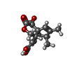  Mass: 276.285 Da / Num. of mol.: 1 / Source method: obtained synthetically / Formula: C15H16O5 Mass: 276.285 Da / Num. of mol.: 1 / Source method: obtained synthetically / Formula: C15H16O5 |
| #4: Water | ChemComp-HOH /  Mass: 18.015 Da / Num. of mol.: 283 / Source method: isolated from a natural source / Formula: H2O Mass: 18.015 Da / Num. of mol.: 283 / Source method: isolated from a natural source / Formula: H2O |
-Experimental details
-Experiment
| Experiment | Method: X-RAY DIFFRACTION / Number of used crystals: 1 |
|---|
- Sample preparation
| Crystal | Density Matthews: 3.35 Å3/Da / Density % sol: 63.26 % / Mosaicity: 0.22 ° |
|---|---|
| Crystal grow | Temperature: 288 K / Method: evaporation / pH: 9 / Details: Bicine, sodium citrate, glycerol |
-Data collection
| Diffraction | Mean temperature: 100 K | ||||||||||||||||||||||||||||||
|---|---|---|---|---|---|---|---|---|---|---|---|---|---|---|---|---|---|---|---|---|---|---|---|---|---|---|---|---|---|---|---|
| Diffraction source | Source: ROTATING ANODE / Type: RIGAKU FR-E SUPERBRIGHT / Wavelength: 1.54179 Å | ||||||||||||||||||||||||||||||
| Detector | Type: RIGAKU SATURN 944+ / Detector: CCD / Date: Oct 31, 2014 / Details: mirrors | ||||||||||||||||||||||||||||||
| Radiation | Protocol: SINGLE WAVELENGTH / Monochromatic (M) / Laue (L): M / Scattering type: x-ray | ||||||||||||||||||||||||||||||
| Radiation wavelength | Wavelength: 1.54179 Å / Relative weight: 1 | ||||||||||||||||||||||||||||||
| Reflection | Resolution: 2.28→45.33 Å / Num. obs: 27950 / % possible obs: 100 % / Redundancy: 7.2 % / Biso Wilson estimate: 20.85 Å2 / CC1/2: 0.983 / Rmerge(I) obs: 0.303 / Net I/σ(I): 6.4 | ||||||||||||||||||||||||||||||
| Reflection shell | Diffraction-ID: 1 / Rejects: _
|
- Processing
| Software |
| ||||||||||||||||||||||||||||||||||||||||||||||||||||||||||||||||||
|---|---|---|---|---|---|---|---|---|---|---|---|---|---|---|---|---|---|---|---|---|---|---|---|---|---|---|---|---|---|---|---|---|---|---|---|---|---|---|---|---|---|---|---|---|---|---|---|---|---|---|---|---|---|---|---|---|---|---|---|---|---|---|---|---|---|---|---|
| Refinement | Method to determine structure: MOLECULAR REPLACEMENT Starting model: 2x9p Resolution: 2.28→45.326 Å / SU ML: 0.23 / Cross valid method: FREE R-VALUE / σ(F): 1.34 / Phase error: 21.72
| ||||||||||||||||||||||||||||||||||||||||||||||||||||||||||||||||||
| Solvent computation | Shrinkage radii: 0.9 Å / VDW probe radii: 1.11 Å | ||||||||||||||||||||||||||||||||||||||||||||||||||||||||||||||||||
| Displacement parameters | Biso max: 103.38 Å2 / Biso mean: 27.1348 Å2 / Biso min: 9.36 Å2 | ||||||||||||||||||||||||||||||||||||||||||||||||||||||||||||||||||
| Refinement step | Cycle: final / Resolution: 2.28→45.326 Å
| ||||||||||||||||||||||||||||||||||||||||||||||||||||||||||||||||||
| Refine LS restraints |
| ||||||||||||||||||||||||||||||||||||||||||||||||||||||||||||||||||
| LS refinement shell | Refine-ID: X-RAY DIFFRACTION / Total num. of bins used: 10 / % reflection obs: 100 %
| ||||||||||||||||||||||||||||||||||||||||||||||||||||||||||||||||||
| Refinement TLS params. | Method: refined / Origin x: 20.8177 Å / Origin y: 26.8278 Å / Origin z: 20.2422 Å
| ||||||||||||||||||||||||||||||||||||||||||||||||||||||||||||||||||
| Refinement TLS group |
|
