 Movie
Movie Controller

[English] 日本語
 Yorodumi
- PDB-5l0q: Crystal structure of the complex between ADAM10 D+C domain and a ... -
+ Open data

- Basic information
| Entry | Database: PDB / ID: 5l0q | ||||||
|---|---|---|---|---|---|---|---|
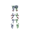
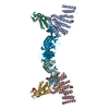
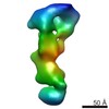
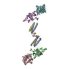
| Title | Crystal structure of the complex between ADAM10 D+C domain and a conformation specific mAb 8C7. | ||||||
 Components Components |
| ||||||
 Keywords Keywords | HYDROLASE/IMMUNE SYSTEM / ADAM protease / mAb / 8C7 / Notch signaling / therapeutic antibody / cancer stem cell / drug resistance / HYDROLASE-IMMUNE SYSTEM complex | ||||||
| Function / homology |  Function and homology information Function and homology informationDegradation of the extracellular matrix / ADAM10 endopeptidase / constitutive protein ectodomain proteolysis / regulation of vasculature development / Regulation of Insulin-like Growth Factor (IGF) transport and uptake by Insulin-like Growth Factor Binding Proteins (IGFBPs) / Post-translational protein phosphorylation / epidermal growth factor receptor ligand maturation / monocyte activation / metalloendopeptidase activity involved in amyloid precursor protein catabolic process / protein catabolic process at postsynapse ...Degradation of the extracellular matrix / ADAM10 endopeptidase / constitutive protein ectodomain proteolysis / regulation of vasculature development / Regulation of Insulin-like Growth Factor (IGF) transport and uptake by Insulin-like Growth Factor Binding Proteins (IGFBPs) / Post-translational protein phosphorylation / epidermal growth factor receptor ligand maturation / monocyte activation / metalloendopeptidase activity involved in amyloid precursor protein catabolic process / protein catabolic process at postsynapse / postsynapse organization / perinuclear endoplasmic reticulum / tetraspanin-enriched microdomain / regulation of Notch signaling pathway / positive regulation of T cell chemotaxis / pore complex assembly / metallodipeptidase activity / negative regulation of cell adhesion / adherens junction organization / positive regulation of tumor necrosis factor-mediated signaling pathway / clathrin-coated vesicle / regulation of postsynapse organization / Neutrophil degranulation / Golgi-associated vesicle / amyloid precursor protein catabolic process / pore complex / regulation of neurotransmitter receptor localization to postsynaptic specialization membrane / membrane protein ectodomain proteolysis / cochlea development / response to tumor necrosis factor / Notch signaling pathway / synaptic membrane / adherens junction / protein processing / metalloendopeptidase activity / SH3 domain binding / metallopeptidase activity / positive regulation of tumor necrosis factor production / in utero embryonic development / positive regulation of cell growth / endopeptidase activity / postsynaptic density / Golgi membrane / axon / positive regulation of cell population proliferation / dendrite / glutamatergic synapse / Golgi apparatus / cell surface / protein homodimerization activity / metal ion binding / plasma membrane Similarity search - Function | ||||||
| Biological species |   Homo sapiens (human) Homo sapiens (human) | ||||||
| Method |  X-RAY DIFFRACTION / SYNCHROTRON / MOLECULAR REPLACEMENT / Resolution: 2.759 Å X-RAY DIFFRACTION / SYNCHROTRON / MOLECULAR REPLACEMENT / Resolution: 2.759 Å | ||||||
 Authors Authors | Xu, K. / Saha, N. / Nikolov, D.B. | ||||||
 Citation Citation | Journal: J.Exp.Med. / Year: 2016 Title: An activated form of ADAM10 is tumor selective and regulates cancer stem-like cells and tumor growth. Authors: Atapattu, L. / Saha, N. / Chheang, C. / Eissman, M.F. / Xu, K. / Vail, M.E. / Hii, L. / Llerena, C. / Liu, Z. / Horvay, K. / Abud, H.E. / Kusebauch, U. / Moritz, R.L. / Ding, B.S. / Cao, Z. ...Authors: Atapattu, L. / Saha, N. / Chheang, C. / Eissman, M.F. / Xu, K. / Vail, M.E. / Hii, L. / Llerena, C. / Liu, Z. / Horvay, K. / Abud, H.E. / Kusebauch, U. / Moritz, R.L. / Ding, B.S. / Cao, Z. / Rafii, S. / Ernst, M. / Scott, A.M. / Nikolov, D.B. / Lackmann, M. / Janes, P.W. | ||||||
| History |
|
- Structure visualization
| Structure viewer | Molecule: MolmilJmol/JSmol |
|---|
- Downloads & links
-Download
| PDBx/mmCIF format | 5l0q.cif.gz | 259.8 KB | Display | PDBx/mmCIF format |
|---|---|---|---|---|
| PDB format | pdb5l0q.ent.gz | 207.1 KB | Display | PDB format |
| PDBx/mmJSON format | 5l0q.json.gz | Tree view | PDBx/mmJSON format | |
| Others |  Other downloads Other downloads |
-Validation report
| Arichive directory | https://data.pdbj.org/pub/pdb/validation_reports/l0/5l0qftp://data.pdbj.org/pub/pdb/validation_reports/l0/5l0q | HTTPS FTP |
|---|
-Related structure data
| Related structure data |  2ao7S S: Starting model for refinement |
|---|---|
| Similar structure data |


-Links
 PDBj
PDBj


- Assembly
| Deposited unit | 
| ||||||||
|---|---|---|---|---|---|---|---|---|---|
| 1 |
| ||||||||
| Unit cell |
|
-Components
-Antibody , 2 types, 4 molecules BECF
| #2: Antibody | Mass: 23712.094 Da / Num. of mol.: 2 Source method: isolated from a genetically manipulated source Source: (gene. exp.) Homo sapiens (human) / Production host: Homo sapiens (human)#3: Antibody | Mass: 23930.732 Da / Num. of mol.: 2 Source method: isolated from a genetically manipulated source Source: (gene. exp.) Homo sapiens (human) / Production host: Homo sapiens (human) |
|---|
-Protein / Sugars , 2 types, 6 molecules AD
| #1: Protein | Mass: 21780.699 Da / Num. of mol.: 2 / Fragment: UNP residues 455-646 Source method: isolated from a genetically manipulated source Source: (gene. exp.) Homo sapiens (human) / References: UniProt: Q10741, ADAM10 endopeptidase#4: Sugar | ChemComp-NAG /  Type: D-saccharide, beta linking / Mass: 221.208 Da / Num. of mol.: 4 Type: D-saccharide, beta linking / Mass: 221.208 Da / Num. of mol.: 4Source method: isolated from a genetically manipulated source Formula: C8H15NO6 |
|---|
-Non-polymers , 3 types, 349 molecules 


| #5: Chemical | ChemComp-SO4 /  Mass: 96.063 Da / Num. of mol.: 13 / Source method: obtained synthetically / Formula: SO4 Mass: 96.063 Da / Num. of mol.: 13 / Source method: obtained synthetically / Formula: SO4#6: Chemical |  Mass: 24.305 Da / Num. of mol.: 2 / Source method: obtained synthetically / Formula: Mg Mass: 24.305 Da / Num. of mol.: 2 / Source method: obtained synthetically / Formula: Mg#7: Water | ChemComp-HOH / | Mass: 18.015 Da / Num. of mol.: 334 / Source method: isolated from a natural source / Formula: H2O |
|---|
-Details
| Has protein modification | Y |
|---|
-Experimental details
-Experiment
| Experiment | Method: X-RAY DIFFRACTION / Number of used crystals: 1 |
|---|
- Sample preparation
| Crystal | Density Matthews: 3.66 Å3/Da / Density % sol: 66.37 % |
|---|---|
| Crystal grow | Temperature: 293 K / Method: vapor diffusion, hanging drop / Details: 0.1M HEPES, 0.2M NaCl, and 1.6 M ammonium sulphate |
-Data collection
| Diffraction | Mean temperature: 100 K | ||||||||||||||||||||||||||||||||||||||||||||||||||||||||||||||||||
|---|---|---|---|---|---|---|---|---|---|---|---|---|---|---|---|---|---|---|---|---|---|---|---|---|---|---|---|---|---|---|---|---|---|---|---|---|---|---|---|---|---|---|---|---|---|---|---|---|---|---|---|---|---|---|---|---|---|---|---|---|---|---|---|---|---|---|---|
| Diffraction source | Source: SYNCHROTRON / Site: APS  / Beamline: 24-ID-C / Wavelength: 0.9792 Å / Beamline: 24-ID-C / Wavelength: 0.9792 Å | ||||||||||||||||||||||||||||||||||||||||||||||||||||||||||||||||||
| Detector | Type: DECTRIS PILATUS 6M-F / Detector: PIXEL / Date: Oct 20, 2013 | ||||||||||||||||||||||||||||||||||||||||||||||||||||||||||||||||||
| Radiation | Protocol: SINGLE WAVELENGTH / Monochromatic (M) / Laue (L): M / Scattering type: x-ray | ||||||||||||||||||||||||||||||||||||||||||||||||||||||||||||||||||
| Radiation wavelength | Wavelength: 0.9792 Å / Relative weight: 1 | ||||||||||||||||||||||||||||||||||||||||||||||||||||||||||||||||||
| Reflection | Resolution: 2.759→125.262 Å / Num. all: 52855 / Num. obs: 52855 / % possible obs: 99.1 % / Redundancy: 4.4 % / Rpim(I) all: 0.071 / Rrim(I) all: 0.155 / Rsym value: 0.125 / Net I/av σ(I): 5.556 / Net I/σ(I): 10.3 / Num. measured all: 234255 | ||||||||||||||||||||||||||||||||||||||||||||||||||||||||||||||||||
| Reflection shell |
|
- Processing
| Software |
| ||||||||||||||||||||||||||||||||||||||||||||||||||||||||||||||||||||||||||||||||||||||||||||||||||||||||||||||||||||||||||||||||||||||||||||
|---|---|---|---|---|---|---|---|---|---|---|---|---|---|---|---|---|---|---|---|---|---|---|---|---|---|---|---|---|---|---|---|---|---|---|---|---|---|---|---|---|---|---|---|---|---|---|---|---|---|---|---|---|---|---|---|---|---|---|---|---|---|---|---|---|---|---|---|---|---|---|---|---|---|---|---|---|---|---|---|---|---|---|---|---|---|---|---|---|---|---|---|---|---|---|---|---|---|---|---|---|---|---|---|---|---|---|---|---|---|---|---|---|---|---|---|---|---|---|---|---|---|---|---|---|---|---|---|---|---|---|---|---|---|---|---|---|---|---|---|---|---|
| Refinement | Method to determine structure: MOLECULAR REPLACEMENT Starting model: 2AO7 Resolution: 2.759→125.262 Å / SU ML: 0.36 / Cross valid method: FREE R-VALUE / σ(F): 1.34 / Phase error: 27.44 / Stereochemistry target values: ML
| ||||||||||||||||||||||||||||||||||||||||||||||||||||||||||||||||||||||||||||||||||||||||||||||||||||||||||||||||||||||||||||||||||||||||||||
| Solvent computation | Shrinkage radii: 0.9 Å / VDW probe radii: 1.11 Å / Solvent model: FLAT BULK SOLVENT MODEL | ||||||||||||||||||||||||||||||||||||||||||||||||||||||||||||||||||||||||||||||||||||||||||||||||||||||||||||||||||||||||||||||||||||||||||||
| Refinement step | Cycle: LAST / Resolution: 2.759→125.262 Å
| ||||||||||||||||||||||||||||||||||||||||||||||||||||||||||||||||||||||||||||||||||||||||||||||||||||||||||||||||||||||||||||||||||||||||||||
| Refine LS restraints |
| ||||||||||||||||||||||||||||||||||||||||||||||||||||||||||||||||||||||||||||||||||||||||||||||||||||||||||||||||||||||||||||||||||||||||||||
| LS refinement shell |
|
