 Movie
Movie Controller


+ Open data

- Basic information
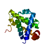
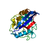
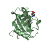
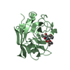
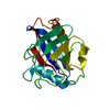
| Entry | Database: PDB / ID: 5kuv | |||||||||||||||
|---|---|---|---|---|---|---|---|---|---|---|---|---|---|---|---|---|
| Title | Human cyclophilin A at 100K, Data set 8 | |||||||||||||||
 Components Components | Peptidyl-prolyl cis-trans isomerase A | |||||||||||||||
 Keywords Keywords | ISOMERASE / Conformational variation / Radiation damage | |||||||||||||||
| Function / homology |  Function and homology information Function and homology informationnegative regulation of protein K48-linked ubiquitination / regulation of apoptotic signaling pathway / cell adhesion molecule production / lipid droplet organization / negative regulation of viral life cycle / heparan sulfate binding / regulation of viral genome replication / virion binding / leukocyte chemotaxis / negative regulation of stress-activated MAPK cascade ...negative regulation of protein K48-linked ubiquitination / regulation of apoptotic signaling pathway / cell adhesion molecule production / lipid droplet organization / negative regulation of viral life cycle / heparan sulfate binding / regulation of viral genome replication / virion binding / leukocyte chemotaxis / negative regulation of stress-activated MAPK cascade / activation of protein kinase B activity / endothelial cell activation / Basigin interactions / protein peptidyl-prolyl isomerization / cyclosporin A binding / Minus-strand DNA synthesis / Plus-strand DNA synthesis / Uncoating of the HIV Virion / Early Phase of HIV Life Cycle / Integration of provirus / APOBEC3G mediated resistance to HIV-1 infection / negative regulation of protein phosphorylation / viral release from host cell / Calcineurin activates NFAT / Binding and entry of HIV virion / negative regulation of protein kinase activity / negative regulation of oxidative stress-induced intrinsic apoptotic signaling pathway / positive regulation of viral genome replication / neutrophil chemotaxis / Gene and protein expression by JAK-STAT signaling after Interleukin-12 stimulation / : / peptidylprolyl isomerase / positive regulation of protein secretion / peptidyl-prolyl cis-trans isomerase activity / Assembly Of The HIV Virion / platelet activation / Budding and maturation of HIV virion / positive regulation of protein phosphorylation / platelet aggregation / integrin binding / neuron differentiation / SARS-CoV-1 activates/modulates innate immune responses / : / Platelet degranulation / protein folding / cellular response to oxidative stress / secretory granule lumen / vesicle / ficolin-1-rich granule lumen / positive regulation of MAPK cascade / focal adhesion / apoptotic process / Neutrophil degranulation / protein-containing complex / : / RNA binding / extracellular exosome / extracellular region / membrane / nucleus / cytoplasm / cytosol Similarity search - Function | |||||||||||||||
| Biological species |  Homo sapiens (human) Homo sapiens (human) | |||||||||||||||
| Method |  X-RAY DIFFRACTION / SYNCHROTRON / MOLECULAR REPLACEMENT / Resolution: 1.7 Å X-RAY DIFFRACTION / SYNCHROTRON / MOLECULAR REPLACEMENT / Resolution: 1.7 Å | |||||||||||||||
 Authors Authors | Russi, S. / Gonzalez, A. / Kenner, L.R. / Keedy, D.A. / Fraser, J.S. / van den Bedem, H. | |||||||||||||||
| Funding support |  United States, 4items United States, 4items
| |||||||||||||||
 Citation Citation | Journal: J Synchrotron Radiat / Year: 2017 Title: Conformational variation of proteins at room temperature is not dominated by radiation damage. Authors: Russi, S. / Gonzalez, A. / Kenner, L.R. / Keedy, D.A. / Fraser, J.S. / van den Bedem, H. | |||||||||||||||
| History |
|
- Structure visualization
| Structure viewer | Molecule: MolmilJmol/JSmol |
|---|
- Downloads & links
-Download
| PDBx/mmCIF format | 5kuv.cif.gz | 61.7 KB | Display | PDBx/mmCIF format |
|---|---|---|---|---|
| PDB format | pdb5kuv.ent.gz | 44.5 KB | Display | PDB format |
| PDBx/mmJSON format | 5kuv.json.gz | Tree view | PDBx/mmJSON format | |
| Others |  Other downloads Other downloads |
-Validation report
| Arichive directory | https://data.pdbj.org/pub/pdb/validation_reports/ku/5kuvftp://data.pdbj.org/pub/pdb/validation_reports/ku/5kuv | HTTPS FTP |
|---|
-Related structure data
| Related structure data |  5kulC  5kunC  5kuoC  5kuqC  5kurC  5kusC  5kuuC  5kuwC  5kuzC  5kv0C  5kv1C  5kv2C  5kv3C  5kv4C  5kv5C  5kv6C  5kv7C 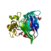 5kvwC  5kvxC  5kvzC  5kw0C  5kw3C  5kw4C  5kw5C  5kw7C 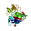 5kw8C  5kxkC  5kxlC  5kxmC 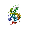 5kxnC  5kxoC  5kxpC  5kxrC  5kxsC  5kxtC  5kxwC 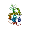 5kxxC  5kxyC  5kxzC  5ky1C  5f66S C: citing same article ( S: Starting model for refinement |
|---|---|
| Similar structure data |


-Links
 PDBj
PDBj


- Assembly
| Deposited unit | 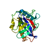
| ||||||||
|---|---|---|---|---|---|---|---|---|---|
| 1 |
| ||||||||
| Unit cell |
|
-Components
| #1: Protein | Mass: 17905.307 Da / Num. of mol.: 1 Source method: isolated from a genetically manipulated source Source: (gene. exp.) Homo sapiens (human) / Gene: PPIA, CYPA / Production host:  |
|---|---|
| #2: Water | ChemComp-HOH /  Mass: 18.015 Da / Num. of mol.: 230 / Source method: isolated from a natural source / Formula: H2O Mass: 18.015 Da / Num. of mol.: 230 / Source method: isolated from a natural source / Formula: H2O |
-Experimental details
-Experiment
| Experiment | Method: X-RAY DIFFRACTION / Number of used crystals: 1 |
|---|
- Sample preparation
| Crystal | Density Matthews: 2.78 Å3/Da / Density % sol: 55.69 % |
|---|---|
| Crystal grow | Temperature: 291 K / Method: vapor diffusion, hanging drop / pH: 7.5 Details: CRYSTALS WERE GROWN BY MIXING EQUAL VOLUMES OF WELL SOLUTION (100 MM HEPES PH 7.5, 23% PEG 3350, 5 MM TCEP) AND PROTEIN (60 MG/ML IN 20 MM HEPES PH 7.5, 100 MM NACL, 0.5 MM TCEP) IN THE HANGING-DROP FORMAT. |
-Data collection
| Diffraction | Mean temperature: 100 K |
|---|---|
| Diffraction source | Source: SYNCHROTRON / Site: SSRL / Beamline: BL7-1 / Wavelength: 1.1 Å |
| Detector | Type: ADSC QUANTUM 315r / Detector: CCD / Date: Apr 17, 2014 |
| Radiation | Protocol: SINGLE WAVELENGTH / Monochromatic (M) / Laue (L): M / Scattering type: x-ray |
| Radiation wavelength | Wavelength: 1.1 Å / Relative weight: 1 |
| Reflection | Resolution: 1.48→38.24 Å / Num. obs: 31607 / % possible obs: 94 % / Redundancy: 2.8 % / Biso Wilson estimate: 18.56 Å2 / CC1/2: 0.998 / Rmerge(I) obs: 0.062 / Rpim(I) all: 0.043 / Rrim(I) all: 0.076 / Net I/σ(I): 12.3 / Num. measured all: 87869 / Scaling rejects: 194 |
| Reflection shell | Resolution: 1.48→1.51 Å / Redundancy: 2.2 % / Rmerge(I) obs: 1.182 / CC1/2: 0.265 / % possible all: 58.6 |
- Processing
| Software |
| ||||||||||||||||||||||||||||||||||||||||||||||||||||||||
|---|---|---|---|---|---|---|---|---|---|---|---|---|---|---|---|---|---|---|---|---|---|---|---|---|---|---|---|---|---|---|---|---|---|---|---|---|---|---|---|---|---|---|---|---|---|---|---|---|---|---|---|---|---|---|---|---|---|
| Refinement | Method to determine structure: MOLECULAR REPLACEMENT Starting model: 5F66 Resolution: 1.7→34.042 Å / SU ML: 0.17 / Cross valid method: FREE R-VALUE / σ(F): 1.34 / Phase error: 20.39
| ||||||||||||||||||||||||||||||||||||||||||||||||||||||||
| Solvent computation | Shrinkage radii: 0.9 Å / VDW probe radii: 1.11 Å | ||||||||||||||||||||||||||||||||||||||||||||||||||||||||
| Displacement parameters | Biso max: 91.12 Å2 / Biso mean: 23.8265 Å2 / Biso min: 10.53 Å2 | ||||||||||||||||||||||||||||||||||||||||||||||||||||||||
| Refinement step | Cycle: final / Resolution: 1.7→34.042 Å
| ||||||||||||||||||||||||||||||||||||||||||||||||||||||||
| Refine LS restraints |
| ||||||||||||||||||||||||||||||||||||||||||||||||||||||||
| LS refinement shell |
|
