 Movie
Movie Controller

[English] 日本語
 Yorodumi
- PDB-5kp9: Structure of Nanoparticle Released from Enveloped Protein Nanoparticle -
+ Open data

- Basic information
| Entry | Database: PDB / ID: 5kp9 | ||||||||||||||||||||||||||||||||||||||||
|---|---|---|---|---|---|---|---|---|---|---|---|---|---|---|---|---|---|---|---|---|---|---|---|---|---|---|---|---|---|---|---|---|---|---|---|---|---|---|---|---|---|
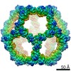
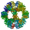
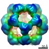
| Title | Structure of Nanoparticle Released from Enveloped Protein Nanoparticle | ||||||||||||||||||||||||||||||||||||||||
 Components Components | EPN-01* Keywords KeywordsSTRUCTURAL PROTEIN / protein design / icosahedral assemblies / cell transduction / enveloped viruses / virus assembly / enveloped protein / nanoparticle | Function / homology |  Function and homology information Function and homology informationD-galactonate catabolic process / 2-dehydro-3-deoxy-6-phosphogalactonate aldolase activity / viral budding via host ESCRT complex / host multivesicular body / viral nucleocapsid / viral translational frameshifting / host cell plasma membrane / host cell nucleus / virion membrane / structural molecule activity ...D-galactonate catabolic process / 2-dehydro-3-deoxy-6-phosphogalactonate aldolase activity / viral budding via host ESCRT complex / host multivesicular body / viral nucleocapsid / viral translational frameshifting / host cell plasma membrane / host cell nucleus / virion membrane / structural molecule activity / RNA binding / zinc ion binding / identical protein binding Similarity search - Function Biological species |   Thermotoga maritima (bacteria) Thermotoga maritima (bacteria) Human immunodeficiency virus type 1 group M subtype B Human immunodeficiency virus type 1 group M subtype BMethod | ELECTRON MICROSCOPY / single particle reconstruction / cryo EM / Resolution: 5.7 Å |  Authors AuthorsVotteler, J. / Ogohara, C. / Yi, S. / Hsia, Y. / Natterman, U. / Belnap, D.M. / King, N.P. / Sundquist, W.I. | Funding support | |  Germany, Germany,  United States, 5items United States, 5items
 CitationJournal: Nature / Year: 2016 CitationJournal: Nature / Year: 2016Title: Designed proteins induce the formation of nanocage-containing extracellular vesicles. Authors: Jörg Votteler / Cassandra Ogohara / Sue Yi / Yang Hsia / Una Nattermann / David M Belnap / Neil P King / Wesley I Sundquist / Abstract: Complex biological processes are often performed by self-organizing nanostructures comprising multiple classes of macromolecules, such as ribosomes (proteins and RNA) or enveloped viruses (proteins, ...Complex biological processes are often performed by self-organizing nanostructures comprising multiple classes of macromolecules, such as ribosomes (proteins and RNA) or enveloped viruses (proteins, nucleic acids and lipids). Approaches have been developed for designing self-assembling structures consisting of either nucleic acids or proteins, but strategies for engineering hybrid biological materials are only beginning to emerge. Here we describe the design of self-assembling protein nanocages that direct their own release from human cells inside small vesicles in a manner that resembles some viruses. We refer to these hybrid biomaterials as 'enveloped protein nanocages' (EPNs). Robust EPN biogenesis requires protein sequence elements that encode three distinct functions: membrane binding, self-assembly, and recruitment of the endosomal sorting complexes required for transport (ESCRT) machinery. A variety of synthetic proteins with these functional elements induce EPN biogenesis, highlighting the modularity and generality of the design strategy. Biochemical analyses and cryo-electron microscopy reveal that one design, EPN-01, comprises small (~100 nm) vesicles containing multiple protein nanocages that closely match the structure of the designed 60-subunit self-assembling scaffold. EPNs that incorporate the vesicular stomatitis viral glycoprotein can fuse with target cells and deliver their contents, thereby transferring cargoes from one cell to another. These results show how proteins can be programmed to direct the formation of hybrid biological materials that perform complex tasks, and establish EPNs as a class of designed, modular, genetically-encoded nanomaterials that can transfer molecules between cells. History |
|
- Structure visualization
| Movie |
Movie viewer |
|---|---|
| Structure viewer | Molecule: MolmilJmol/JSmol |
- Downloads & links
-Download
| PDBx/mmCIF format | 5kp9.cif.gz | 52.8 KB | Display | PDBx/mmCIF format |
|---|---|---|---|---|
| PDB format | pdb5kp9.ent.gz | 35 KB | Display | PDB format |
| PDBx/mmJSON format | 5kp9.json.gz | Tree view | PDBx/mmJSON format | |
| Others |  Other downloads Other downloads |
-Validation report
| Arichive directory | https://data.pdbj.org/pub/pdb/validation_reports/kp/5kp9ftp://data.pdbj.org/pub/pdb/validation_reports/kp/5kp9 | HTTPS FTP |
|---|
-Related structure data
| Related structure data | 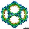 8278MC M: map data used to model this data C: citing same article ( |
|---|---|
| Similar structure data |

-Links
 PDBj
PDBj


- Assembly
| Deposited unit | 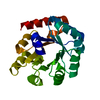
|
|---|---|
| 1 | x 60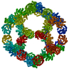
|
| 2 |
|
| 3 | x 5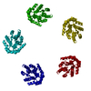
|
| 4 | x 6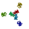
|
| 5 | 
|
| Symmetry | Point symmetry: (Schoenflies symbol: I (icosahedral)) |
-Components
| #1: Protein | Mass: 30304.920 Da / Num. of mol.: 1 Source method: isolated from a genetically manipulated source Source: (gene. exp.) Thermotoga maritima (bacteria), (gene. exp.) Human immunodeficiency virus type 1 group M subtype B (isolate BH10)Gene: TM_0066, gag / Strain: isolate BH10 / Cell line (production host): HEK293T / Production host:  Homo sapiens (human) / References: UniProt: Q9WXS1, UniProt: P03347 Homo sapiens (human) / References: UniProt: Q9WXS1, UniProt: P03347 |
|---|---|
| Has protein modification | Y |
-Experimental details
-Experiment
| Experiment | Method: ELECTRON MICROSCOPY |
|---|---|
| EM experiment | Aggregation state: PARTICLE / 3D reconstruction method: single particle reconstruction |
- Sample preparation
| Component | Name: EPN-01* / Type: COMPLEX / Entity ID: all / Source: RECOMBINANT |
|---|---|
| Source (natural) | Organism: Thermotoga maritima (bacteria) |
| Source (recombinant) | Organism: Homo sapiens (human) / Cell: HEK293T / Plasmid: EPN-01* |
| Buffer solution | pH: 7.5 / Details: Phosphate-buffered saline |
| Specimen | Conc.: 0.2 mg/ml / Embedding applied: NO / Shadowing applied: NO / Staining applied: NO / Vitrification applied: YES Details: EPN nanoparticles released from vesicles by detergent treatment (0.75% CHAPS) |
| Specimen support | Grid material: COPPER / Grid type: Quantifoil R2/2 |
| Vitrification | Instrument: FEI VITROBOT MARK II / Cryogen name: ETHANE / Humidity: 80 % / Chamber temperature: 277 K / Details: 11 second blot, 0 mm offset |
- Electron microscopy imaging
| Experimental equipment |  Model: Tecnai F20 / Image courtesy: FEI Company |
|---|---|
| Microscopy | Model: FEI TECNAI F20 |
| Electron gun | Electron source:  FIELD EMISSION GUN / Accelerating voltage: 200 kV / Illumination mode: FLOOD BEAM FIELD EMISSION GUN / Accelerating voltage: 200 kV / Illumination mode: FLOOD BEAM |
| Electron lens | Mode: BRIGHT FIELD / Calibrated magnification: 41911 X / Calibrated defocus min: 700 nm / Calibrated defocus max: 3300 nm / Cs: 2 mm / Alignment procedure: COMA FREE |
| Specimen holder | Cryogen: NITROGEN Specimen holder model: GATAN 626 SINGLE TILT LIQUID NITROGEN CRYO TRANSFER HOLDER |
| Image recording | Electron dose: 2 e/Å2 / Detector mode: SUPER-RESOLUTION / Film or detector model: GATAN K2 SUMMIT (4k x 4k) / Details: Electron dose at specimen was not recorded. |
| Image scans | Sampling size: 2.5 µm / Width: 7420 / Height: 7676 / Movie frames/image: 60 |
- Processing
| EM software |
| |||||||||||||||||||||||||||||||||||||||||||||
|---|---|---|---|---|---|---|---|---|---|---|---|---|---|---|---|---|---|---|---|---|---|---|---|---|---|---|---|---|---|---|---|---|---|---|---|---|---|---|---|---|---|---|---|---|---|---|
| CTF correction | Type: PHASE FLIPPING AND AMPLITUDE CORRECTION | |||||||||||||||||||||||||||||||||||||||||||||
| Particle selection | Num. of particles selected: 9177 | |||||||||||||||||||||||||||||||||||||||||||||
| Symmetry | Point symmetry: I (icosahedral) | |||||||||||||||||||||||||||||||||||||||||||||
| 3D reconstruction | Resolution: 5.7 Å / Resolution method: FSC 0.143 CUT-OFF / Num. of particles: 8573 / Algorithm: FOURIER SPACE / Num. of class averages: 10 / Symmetry type: POINT | |||||||||||||||||||||||||||||||||||||||||||||
| Atomic model building | Protocol: RIGID BODY FIT |
