 Movie
Movie Controller


+ Open data

- Basic information
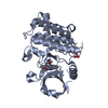
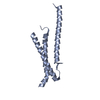
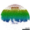
| Entry | Database: PDB / ID: 5kmg | ||||||||||||
|---|---|---|---|---|---|---|---|---|---|---|---|---|---|
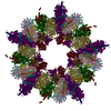
| Title | Near-atomic cryo-EM structure of PRC1 bound to the microtubule | ||||||||||||
 Components Components |
| ||||||||||||
 Keywords Keywords | STRUCTURAL PROTEIN / cytoskeleton / mitosis / microtubule / microtubule-associated protein | ||||||||||||
| Function / homology |  Function and homology information Function and homology informationcontractile ring / mitotic spindle midzone / mitotic spindle elongation / mitotic spindle midzone assembly / RHO GTPases activate CIT / Microtubule-dependent trafficking of connexons from Golgi to the plasma membrane / Resolution of Sister Chromatid Cohesion / Hedgehog 'off' state / Cilium Assembly / Intraflagellar transport ...contractile ring / mitotic spindle midzone / mitotic spindle elongation / mitotic spindle midzone assembly / RHO GTPases activate CIT / Microtubule-dependent trafficking of connexons from Golgi to the plasma membrane / Resolution of Sister Chromatid Cohesion / Hedgehog 'off' state / Cilium Assembly / Intraflagellar transport / COPI-dependent Golgi-to-ER retrograde traffic / Mitotic Prometaphase / Carboxyterminal post-translational modifications of tubulin / RHOH GTPase cycle / EML4 and NUDC in mitotic spindle formation / Sealing of the nuclear envelope (NE) by ESCRT-III / Kinesins / PKR-mediated signaling / Separation of Sister Chromatids / The role of GTSE1 in G2/M progression after G2 checkpoint / Aggrephagy / RHO GTPases activate IQGAPs / RHO GTPases Activate Formins / HSP90 chaperone cycle for steroid hormone receptors (SHR) in the presence of ligand / MHC class II antigen presentation / Recruitment of NuMA to mitotic centrosomes / COPI-mediated anterograde transport / kinesin binding / intercellular bridge / regulation of cytokinesis / spindle microtubule / microtubule cytoskeleton organization / structural constituent of cytoskeleton / neuron migration / spindle / spindle pole / mitotic cell cycle / microtubule cytoskeleton / chromosome / midbody / microtubule binding / microtubule / Hydrolases; Acting on acid anhydrides; Acting on GTP to facilitate cellular and subcellular movement / cell division / GTPase activity / positive regulation of cell population proliferation / protein kinase binding / GTP binding / nucleoplasm / metal ion binding / identical protein binding / nucleus / plasma membrane / cytosol / cytoplasm Similarity search - Function | ||||||||||||
| Biological species |  Homo sapiens (human) Homo sapiens (human) | ||||||||||||
| Method | ELECTRON MICROSCOPY / helical reconstruction / cryo EM / Resolution: 3.5 Å | ||||||||||||
 Authors Authors | Kellogg, E.H. / Howes, S. / Ti, S.-C. / Ramirez-Aportela, E. / Kapoor, T.M. / Chacon, P. / Nogales, E. | ||||||||||||
| Funding support |  United States, United States,  Spain, 3items Spain, 3items
| ||||||||||||
 Citation Citation | Journal: Proc Natl Acad Sci U S A / Year: 2016 Title: Near-atomic cryo-EM structure of PRC1 bound to the microtubule. Authors: Elizabeth H Kellogg / Stuart Howes / Shih-Chieh Ti / Erney Ramírez-Aportela / Tarun M Kapoor / Pablo Chacón / Eva Nogales / Abstract: Proteins that associate with microtubules (MTs) are crucial to generate MT arrays and establish different cellular architectures. One example is PRC1 (protein regulator of cytokinesis 1), which cross- ...Proteins that associate with microtubules (MTs) are crucial to generate MT arrays and establish different cellular architectures. One example is PRC1 (protein regulator of cytokinesis 1), which cross-links antiparallel MTs and is essential for the completion of mitosis and cytokinesis. Here we describe a 4-Å-resolution cryo-EM structure of monomeric PRC1 bound to MTs. Residues in the spectrin domain of PRC1 contacting the MT are highly conserved and interact with the same pocket recognized by kinesin. We additionally found that PRC1 promotes MT assembly even in the presence of the MT stabilizer taxol. Interestingly, the angle of the spectrin domain on the MT surface corresponds to the previously observed cross-bridge angle between MTs cross-linked by full-length, dimeric PRC1. This finding, together with molecular dynamic simulations describing the intrinsic flexibility of PRC1, suggests that the MT-spectrin domain interface determines the geometry of the MT arrays cross-linked by PRC1. | ||||||||||||
| History |
|
- Structure visualization
| Movie |
Movie viewer |
|---|---|
| Structure viewer | Molecule: MolmilJmol/JSmol |
- Downloads & links
-Download
| PDBx/mmCIF format | 5kmg.cif.gz | 205.2 KB | Display | PDBx/mmCIF format |
|---|---|---|---|---|
| PDB format | pdb5kmg.ent.gz | 158.3 KB | Display | PDB format |
| PDBx/mmJSON format | 5kmg.json.gz | Tree view | PDBx/mmJSON format | |
| Others |  Other downloads Other downloads |
-Validation report
| Arichive directory | https://data.pdbj.org/pub/pdb/validation_reports/km/5kmgftp://data.pdbj.org/pub/pdb/validation_reports/km/5kmg | HTTPS FTP |
|---|
-Related structure data
| Related structure data | 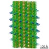 8266MC M: map data used to model this data C: citing same article ( |
|---|---|
| Similar structure data |
-Links
 PDBj
PDBj


- Assembly
| Deposited unit | 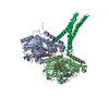
|
|---|---|
| 1 | x 14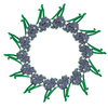
|
| 2 |
|
| 3 | 
|
| Symmetry | Helical symmetry: (Circular symmetry: 1 / Dyad axis: no / N subunits divisor: 1 / Num. of operations: 14 / Rise per n subunits: 8.57 Å / Rotation per n subunits: -25.76 °) |
-Components
-Protein , 3 types, 3 molecules ABP
| #1: Protein | Mass: 49095.438 Da / Num. of mol.: 1 / Source method: isolated from a natural source / Source: (natural) |
|---|---|
| #2: Protein | Mass: 48299.293 Da / Num. of mol.: 1 / Source method: isolated from a natural source / Source: (natural) |
| #3: Protein | Mass: 15693.940 Da / Num. of mol.: 1 / Fragment: UNP residues 341-464 Source method: isolated from a genetically manipulated source Source: (gene. exp.) Homo sapiens (human) / Gene: PRC1 / Production host:  |
-Non-polymers , 4 types, 4 molecules 


| #4: Chemical | ChemComp-GTP /  Mass: 523.180 Da / Num. of mol.: 1 / Source method: obtained synthetically / Formula: C10H16N5O14P3 / Comment: GTP, energy-carrying molecule*YM Mass: 523.180 Da / Num. of mol.: 1 / Source method: obtained synthetically / Formula: C10H16N5O14P3 / Comment: GTP, energy-carrying molecule*YM |
|---|---|
| #5: Chemical | ChemComp-MG /  Mass: 24.305 Da / Num. of mol.: 1 / Source method: obtained synthetically / Formula: Mg Mass: 24.305 Da / Num. of mol.: 1 / Source method: obtained synthetically / Formula: Mg |
| #6: Chemical | ChemComp-GDP /  Type: RNA linking / Mass: 443.201 Da / Num. of mol.: 1 / Source method: obtained synthetically / Formula: C10H15N5O11P2 / Comment: GDP, energy-carrying molecule*YM Type: RNA linking / Mass: 443.201 Da / Num. of mol.: 1 / Source method: obtained synthetically / Formula: C10H15N5O11P2 / Comment: GDP, energy-carrying molecule*YM |
| #7: Chemical | ChemComp-POU /  Mass: 548.663 Da / Num. of mol.: 1 / Source method: obtained synthetically / Formula: C27H48O11 Mass: 548.663 Da / Num. of mol.: 1 / Source method: obtained synthetically / Formula: C27H48O11 |
-Experimental details
-Experiment
| Experiment | Method: ELECTRON MICROSCOPY |
|---|---|
| EM experiment | Aggregation state: HELICAL ARRAY / 3D reconstruction method: helical reconstruction |
- Sample preparation
| Component | Name: Ternary complex of PRC1 fragment (PRC1-SC), alpha-tubulin, and beta-tubulin. Type: COMPLEX Details: The PRC1 fragment referred to as PRC1-SC contains the spectrin domain and the unstructured C-terminal domain, spanning (337-620) Entity ID: #1-#3 / Source: MULTIPLE SOURCES | ||||||||||||||||||||||||||||||
|---|---|---|---|---|---|---|---|---|---|---|---|---|---|---|---|---|---|---|---|---|---|---|---|---|---|---|---|---|---|---|---|
| Buffer solution | pH: 6.8 | ||||||||||||||||||||||||||||||
| Buffer component |
| ||||||||||||||||||||||||||||||
| Specimen | Conc.: 0.4 mg/ml / Embedding applied: NO / Shadowing applied: NO / Staining applied: NO / Vitrification applied: YES | ||||||||||||||||||||||||||||||
| Specimen support | Grid material: COPPER / Grid mesh size: 400 divisions/in. / Grid type: protochips CF-1.2/1.3 | ||||||||||||||||||||||||||||||
| Vitrification | Instrument: FEI VITROBOT MARK IV / Cryogen name: ETHANE / Humidity: 100 % / Chamber temperature: 277.15 K / Details: blot for 4 seconds, blot force 10 |
- Electron microscopy imaging
| Microscopy | Model: FEI TITAN |
|---|---|
| Electron gun | Electron source:  FIELD EMISSION GUN / Accelerating voltage: 300 kV / Illumination mode: FLOOD BEAM FIELD EMISSION GUN / Accelerating voltage: 300 kV / Illumination mode: FLOOD BEAM |
| Electron lens | Mode: BRIGHT FIELD / Nominal magnification: 27500 X / Nominal defocus max: 2500 nm / Nominal defocus min: 1000 nm / Calibrated defocus min: 1100 nm / Calibrated defocus max: 3800 nm / Cs: 2.7 mm / C2 aperture diameter: 50 µm / Alignment procedure: COMA FREE |
| Specimen holder | Cryogen: NITROGEN Specimen holder model: GATAN 626 SINGLE TILT LIQUID NITROGEN CRYO TRANSFER HOLDER Temperature (max): 93 K / Temperature (min): 93 K |
| Image recording | Average exposure time: 0.3 sec. / Electron dose: 8 e/Å2 / Detector mode: COUNTING / Film or detector model: GATAN K2 SUMMIT (4k x 4k) / Num. of grids imaged: 1 / Num. of real images: 312 |
| Image scans | Sampling size: 5 µm / Width: 3710 / Height: 3838 / Movie frames/image: 20 / Used frames/image: 1-20 |
- Processing
| EM software |
| |||||||||||||||||||||||||||||||||||||||||||||||||||||||
|---|---|---|---|---|---|---|---|---|---|---|---|---|---|---|---|---|---|---|---|---|---|---|---|---|---|---|---|---|---|---|---|---|---|---|---|---|---|---|---|---|---|---|---|---|---|---|---|---|---|---|---|---|---|---|---|---|
| CTF correction | Type: PHASE FLIPPING AND AMPLITUDE CORRECTION | |||||||||||||||||||||||||||||||||||||||||||||||||||||||
| Helical symmerty | Angular rotation/subunit: -25.76 ° / Axial rise/subunit: 8.67 Å / Axial symmetry: C1 | |||||||||||||||||||||||||||||||||||||||||||||||||||||||
| Particle selection | Num. of particles selected: 27275 Details: manually selected microtubule filaments using manualpicker.py | |||||||||||||||||||||||||||||||||||||||||||||||||||||||
| 3D reconstruction | Resolution: 3.5 Å / Resolution method: FSC 0.143 CUT-OFF / Num. of particles: 17069 / Algorithm: FOURIER SPACE / Symmetry type: HELICAL | |||||||||||||||||||||||||||||||||||||||||||||||||||||||
| Atomic model building | Protocol: FLEXIBLE FIT / Space: REAL / Target criteria: correlation coefficient | |||||||||||||||||||||||||||||||||||||||||||||||||||||||
| Atomic model building | 3D fitting-ID: 1 / Pdb chain-ID: A / Source name: PDB / Type: experimental model
|

