X-RAY DIFFRACTION / MOLECULAR REPLACEMENT / Resolution: 1.7 Å
Model details
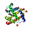
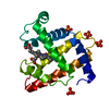
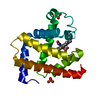
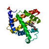
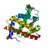
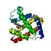
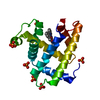
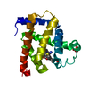
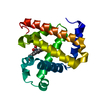
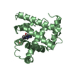
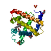
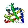
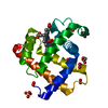
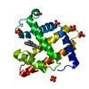
This stable porphyrin-Fe(?Ea)-nitrosoalkane complex was obtained from the reaction of sperm whale ...This stable porphyrin-Fe(?Ea)-nitrosoalkane complex was obtained from the reaction of sperm whale myoglobin ferric H64A and N-hydroxyamphetamine.
Resolution: 1.7→29.36 Å / SU B: 1.329 / SU ML: 0.045 / Cross valid method: THROUGHOUT / σ(F): 0 / ESU R: 0.079 / ESU R Free: 0.08 / Details: HYDROGENS HAVE BEEN ADDED IN THE RIDING
Rfactor
Num. reflection
% reflection
Selection details
Rfree
0.177
1206
5.1 %
RANDOM
Rwork
0.148
-
-
-
obs
0.149
22261
99.9 %
-
Displacement parameters
Biso mean: 16.64 Å2
Baniso -1
Baniso -2
Baniso -3
1-
0.03 Å2
0.01 Å2
0 Å2
2-
-
0.03 Å2
0 Å2
3-
-
-
-0.1 Å2
Refinement step
Cycle: LAST / Resolution: 1.7→29.36 Å
Protein
Nucleic acid
Ligand
Solvent
Total
Num. atoms
1211
0
105
192
1508
+
About Yorodumi
-
News
-
Feb 9, 2022. New format data for meta-information of EMDB entries
New format data for meta-information of EMDB entries
Version 3 of the EMDB header file is now the official format.
The previous official version 1.9 will be removed from the archive.
In the structure databanks used in Yorodumi, some data are registered as the other names, "COVID-19 virus" and "2019-nCoV". Here are the details of the virus and the list of structure data.
Jan 31, 2019. EMDB accession codes are about to change! (news from PDBe EMDB page)
EMDB accession codes are about to change! (news from PDBe EMDB page)
The allocation of 4 digits for EMDB accession codes will soon come to an end. Whilst these codes will remain in use, new EMDB accession codes will include an additional digit and will expand incrementally as the available range of codes is exhausted. The current 4-digit format prefixed with “EMD-” (i.e. EMD-XXXX) will advance to a 5-digit format (i.e. EMD-XXXXX), and so on. It is currently estimated that the 4-digit codes will be depleted around Spring 2019, at which point the 5-digit format will come into force.
The EM Navigator/Yorodumi systems omit the EMD- prefix.
Related info.:Q: What is EMD? / ID/Accession-code notation in Yorodumi/EM Navigator
Yorodumi is a browser for structure data from EMDB, PDB, SASBDB, etc.
This page is also the successor to EM Navigator detail page, and also detail information page/front-end page for Omokage search.
The word "yorodu" (or yorozu) is an old Japanese word meaning "ten thousand". "mi" (miru) is to see.
Related info.:EMDB / PDB / SASBDB / Comparison of 3 databanks / Yorodumi Search / Aug 31, 2016. New EM Navigator & Yorodumi / Yorodumi Papers / Jmol/JSmol / Function and homology information / Changes in new EM Navigator and Yorodumi
 Movie
Movie Controller


 Open data
 Basic information
 Components
 Keywords
 Function and homology information
 X-RAY DIFFRACTION /
  Authors
 United States, 1items
  Citation
 Structure visualization
 Downloads & links
 Other downloads
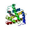


 PDBj
PDBj


 Assembly


 Mass: 616.487 Da / Num. of mol.: 1 / Source method: obtained synthetically / Formula: C34H32FeN4O4
 Mass: 96.063 Da / Num. of mol.: 4 / Source method: obtained synthetically / Formula: SO4
 Mass: 149.190 Da / Num. of mol.: 1 / Source method: obtained synthetically / Formula: C9H11NO
 Mass: 92.094 Da / Num. of mol.: 5 / Source method: obtained synthetically / Formula: C3H8O3
 Sample preparation
 Processing