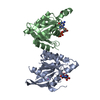
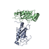
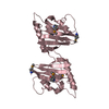
CAMP-dependent protein kinase regulatory subunit, putative
Keywords
TRANSFERASE / Plasmodium falciparum / PKA / protein kinase A / cAMP / CBD / cyclic nucleotide binding / CNB / regulatory domain / R
Function / homology
Function and homology information
PKA activation / PKA activation in glucagon signalling / DARPP-32 events / CREB1 phosphorylation through the activation of Adenylate Cyclase / Hedgehog 'off' state / GPER1 signaling / High laminar flow shear stress activates signaling by PIEZO1 and PECAM1:CDH5:KDR in endothelial cells / Vasopressin regulates renal water homeostasis via Aquaporins / cAMP-dependent protein kinase regulator activity / cAMP-dependent protein kinase inhibitor activity ...PKA activation / PKA activation in glucagon signalling / DARPP-32 events / CREB1 phosphorylation through the activation of Adenylate Cyclase / Hedgehog 'off' state / GPER1 signaling / High laminar flow shear stress activates signaling by PIEZO1 and PECAM1:CDH5:KDR in endothelial cells / Vasopressin regulates renal water homeostasis via Aquaporins / cAMP-dependent protein kinase regulator activity / cAMP-dependent protein kinase inhibitor activity / cAMP-dependent protein kinase / cAMP-dependent protein kinase activity / cAMP-dependent protein kinase complex / protein kinase A catalytic subunit binding / cAMP binding / adenylate cyclase-activating G protein-coupled receptor signaling pathway / protein-containing complex / cytosol Similarity search - Function
Mass: 18.015 Da / Num. of mol.: 36 / Source method: isolated from a natural source / Formula: H2O
-
Experimental details
-
Experiment
Experiment
Method: X-RAY DIFFRACTION / Number of used crystals: 1
-
Sample preparation
Crystal
Density Matthews: 3.07 Å3/Da / Density % sol: 59.97 %
Crystal grow
Temperature: 277 K / Method: vapor diffusion, hanging drop / pH: 7.5 Details: Protein at 12 mg/mL were set in a 1:1 ratio against a reservoir consisting of 20% w/v Polyethylene glycol 3350, 0.1M NaF and 0.1M 1,3-bispropane pH 7.5
-
Data collection
Diffraction
Mean temperature: 100 K
Diffraction source
Source: SYNCHROTRON / Site: Australian Synchrotron / Beamline: MX1 / Wavelength: 0.9537 Å
Resolution: 2→46.458 Å / SU ML: 0.27 / Cross valid method: FREE R-VALUE / σ(F): 1.34 / Phase error: 35.1 / Stereochemistry target values: ML Details: DUE TO N-TERMINAL DISORDER THE IDENTITY OF RESIDUES SER-149, VAL-150, SER-151, ALA-152 IS ONLY TENTATIVE.
Rfactor
Num. reflection
% reflection
Rfree
0.2854
2951
5.06 %
Rwork
0.2454
-
-
obs
0.2474
58355
99.91 %
Solvent computation
Shrinkage radii: 0.9 Å / VDW probe radii: 1.11 Å / Solvent model: FLAT BULK SOLVENT MODEL
Refinement step
Cycle: LAST / Resolution: 2→46.458 Å
Protein
Nucleic acid
Ligand
Solvent
Total
Num. atoms
4535
0
88
36
4659
Refine LS restraints
Refine-ID
Type
Dev ideal
Number
X-RAY DIFFRACTION
f_bond_d
0.009
4703
X-RAY DIFFRACTION
f_angle_d
1.256
6340
X-RAY DIFFRACTION
f_dihedral_angle_d
17.709
1792
X-RAY DIFFRACTION
f_chiral_restr
0.051
712
X-RAY DIFFRACTION
f_plane_restr
0.004
791
LS refinement shell
Resolution (Å)
Rfactor Rfree
Num. reflection Rfree
Rfactor Rwork
Num. reflection Rwork
Refine-ID
% reflection obs (%)
2-2.0328
0.3776
140
0.3596
2624
X-RAY DIFFRACTION
100
2.0328-2.0679
0.362
145
0.3505
2579
X-RAY DIFFRACTION
100
2.0679-2.1055
0.3365
133
0.3257
2593
X-RAY DIFFRACTION
100
2.1055-2.146
0.3078
128
0.3096
2605
X-RAY DIFFRACTION
100
2.146-2.1898
0.3607
123
0.3092
2643
X-RAY DIFFRACTION
100
2.1898-2.2374
0.3257
143
0.2938
2582
X-RAY DIFFRACTION
100
2.2374-2.2894
0.3226
130
0.289
2646
X-RAY DIFFRACTION
100
2.2894-2.3467
0.3001
138
0.285
2604
X-RAY DIFFRACTION
100
2.3467-2.4101
0.3259
150
0.2728
2610
X-RAY DIFFRACTION
100
2.4101-2.481
0.319
136
0.2804
2627
X-RAY DIFFRACTION
100
2.481-2.5611
0.3493
148
0.2847
2613
X-RAY DIFFRACTION
100
2.5611-2.6526
0.3104
149
0.2829
2617
X-RAY DIFFRACTION
100
2.6526-2.7588
0.3334
148
0.2712
2602
X-RAY DIFFRACTION
100
2.7588-2.8844
0.3019
147
0.2767
2641
X-RAY DIFFRACTION
100
2.8844-3.0364
0.3136
147
0.2757
2636
X-RAY DIFFRACTION
100
3.0364-3.2266
0.2838
146
0.2782
2622
X-RAY DIFFRACTION
100
3.2266-3.4757
0.3365
149
0.2485
2648
X-RAY DIFFRACTION
100
3.4757-3.8253
0.2482
140
0.2253
2650
X-RAY DIFFRACTION
100
3.8253-4.3784
0.263
131
0.1984
2713
X-RAY DIFFRACTION
100
4.3784-5.515
0.2056
137
0.2008
2712
X-RAY DIFFRACTION
100
5.515-46.471
0.285
143
0.2236
2837
X-RAY DIFFRACTION
99
Refinement TLS params.
Method: refined / Refine-ID: X-RAY DIFFRACTION
ID
L11 (°2)
L12 (°2)
L13 (°2)
L22 (°2)
L23 (°2)
L33 (°2)
S11 (Å °)
S12 (Å °)
S13 (Å °)
S21 (Å °)
S22 (Å °)
S23 (Å °)
S31 (Å °)
S32 (Å °)
S33 (Å °)
T11 (Å2)
T12 (Å2)
T13 (Å2)
T22 (Å2)
T23 (Å2)
T33 (Å2)
Origin x (Å)
Origin y (Å)
Origin z (Å)
1
4.2377
1.1814
0.9727
7.0288
-2.7422
5.5193
0.0731
0.3502
0.0944
-0.2485
-0.6089
-0.5505
-0.2391
0.4496
0.2912
0.3333
0.129
-0.0329
0.3212
0.1258
0.4044
80.0133
67.6988
101.4679
2
2.9433
-3.5948
1.0769
8.6017
-4.4177
3.457
0.5574
0.854
0.3279
-0.5622
-0.7018
-0.547
0.3166
0.6577
0.2099
0.473
0.1747
0.0784
0.5064
0.2028
0.4827
73.0913
80.2792
86.7162
3
4.5513
-2.5775
0.5178
8.3715
-1.8062
3.1484
-0.0944
-0.3176
0.0914
0.6345
0.5428
0.2535
-0.1683
0.046
-0.2257
0.353
0.0765
0.1183
0.4118
0.2117
0.3586
61.4523
96.5139
90.0731
4
5.3096
2.297
-1.039
5.1813
-2.0397
6.6008
-0.1762
-0.4439
-0.5475
-0.2824
-0.8129
-0.6512
0.3154
0.8805
0.5419
0.4321
0.1874
0.0302
0.6037
0.365
0.5729
40.9246
16.1778
49.4848
5
3.7849
-3.5805
0.889
7.7837
-3.4554
1.9698
-0.2864
-0.8728
-0.3039
0.236
0.7765
0.0509
0.0602
-0.3171
-0.0729
0.3691
0.0348
0.1194
0.7391
0.3525
0.4692
25.3765
39.7576
36.7109
Refinement TLS group
ID
Refine-ID
Refine TLS-ID
Selection details
1
X-RAY DIFFRACTION
1
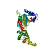
chain 'A' and (resid149through291 )
2
X-RAY DIFFRACTION
2
chain 'A' and (resid292through331 )
3
X-RAY DIFFRACTION
3
chain 'A' and (resid332through441 )
4
X-RAY DIFFRACTION
4
chain 'B' and (resid149through291 )
5
X-RAY DIFFRACTION
5
chain 'B' and (resid292through441 )
+
About Yorodumi
-
News
-
Feb 9, 2022. New format data for meta-information of EMDB entries
New format data for meta-information of EMDB entries
Version 3 of the EMDB header file is now the official format.
The previous official version 1.9 will be removed from the archive.
In the structure databanks used in Yorodumi, some data are registered as the other names, "COVID-19 virus" and "2019-nCoV". Here are the details of the virus and the list of structure data.
Jan 31, 2019. EMDB accession codes are about to change! (news from PDBe EMDB page)
EMDB accession codes are about to change! (news from PDBe EMDB page)
The allocation of 4 digits for EMDB accession codes will soon come to an end. Whilst these codes will remain in use, new EMDB accession codes will include an additional digit and will expand incrementally as the available range of codes is exhausted. The current 4-digit format prefixed with “EMD-” (i.e. EMD-XXXX) will advance to a 5-digit format (i.e. EMD-XXXXX), and so on. It is currently estimated that the 4-digit codes will be depleted around Spring 2019, at which point the 5-digit format will come into force.
The EM Navigator/Yorodumi systems omit the EMD- prefix.
Related info.:Q: What is EMD? / ID/Accession-code notation in Yorodumi/EM Navigator
Yorodumi is a browser for structure data from EMDB, PDB, SASBDB, etc.
This page is also the successor to EM Navigator detail page, and also detail information page/front-end page for Omokage search.
The word "yorodu" (or yorozu) is an old Japanese word meaning "ten thousand". "mi" (miru) is to see.
Related info.:EMDB / PDB / SASBDB / Comparison of 3 databanks / Yorodumi Search / Aug 31, 2016. New EM Navigator & Yorodumi / Yorodumi Papers / Jmol/JSmol / Function and homology information / Changes in new EM Navigator and Yorodumi
 Movie
Movie Controller


 Open data
 Basic information
 Components
 Keywords
 Function and homology information
 X-RAY DIFFRACTION /
  Authors
 Australia, 1items
  Citation
 Structure visualization
 Downloads & links
 Other downloads


 PDBj
PDBj


 Assembly


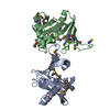
 Mass: 329.206 Da / Num. of mol.: 4 / Source method: obtained synthetically / Formula: C10H12N5O6P
 Mass: 18.015 Da / Num. of mol.: 36 / Source method: isolated from a natural source / Formula: H2O
 Sample preparation
 Processing