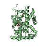
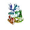
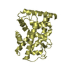
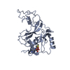
[English] 日本語
Yorodumi
- PDB-5k13: Crystal structure of the RAR alpha ligand-binding domain in compl... -

+
Open data


ID or keywords:

Loading...

-
Basic information

Entry
Database: PDB / ID: 5k13
TitleCrystal structure of the RAR alpha ligand-binding domain in complex with an antagonist
ComponentsRetinoic acid receptor alpha
KeywordsTRANSCRIPTION / NHR ligand-binding domain / antagonist
Function / homology
Function and homology information


Sertoli cell fate commitment / positive regulation of binding / trachea cartilage development / ventricular cardiac muscle cell differentiation / embryonic camera-type eye development / chondroblast differentiation / glandular epithelial cell development / negative regulation of granulocyte differentiation / protein kinase B binding / cellular response to corticotropin-releasing hormone stimulus ...Sertoli cell fate commitment / positive regulation of binding / trachea cartilage development / ventricular cardiac muscle cell differentiation / embryonic camera-type eye development / chondroblast differentiation / glandular epithelial cell development / negative regulation of granulocyte differentiation / protein kinase B binding / cellular response to corticotropin-releasing hormone stimulus / growth plate cartilage development / positive regulation of T-helper 2 cell differentiation / prostate gland development / negative regulation of cartilage development / retinoic acid-responsive element binding / regulation of hematopoietic progenitor cell differentiation / positive regulation of interleukin-13 production / positive regulation of interleukin-5 production / nuclear glucocorticoid receptor binding / retinoic acid binding / response to vitamin A / apoptotic cell clearance / limb development / regulation of myelination / protein kinase A binding / ureteric bud development / Signaling by Retinoic Acid / DNA-binding transcription repressor activity / heterocyclic compound binding / positive regulation of interleukin-4 production / face development / alpha-actinin binding / germ cell development / negative regulation of type II interferon production / negative regulation of tumor necrosis factor production / retinoic acid receptor signaling pathway / cellular response to estrogen stimulus / positive regulation of cell cycle / cellular response to retinoic acid / response to retinoic acid / positive regulation of neuron differentiation / hormone-mediated signaling pathway / mRNA regulatory element binding translation repressor activity / negative regulation of miRNA transcription / liver development / response to cytokine / neural tube closure / female pregnancy / hippocampus development / RNA polymerase II transcription regulatory region sequence-specific DNA binding / SUMOylation of intracellular receptors / mRNA transcription by RNA polymerase II / regulation of synaptic plasticity / multicellular organism growth / chromatin DNA binding / mRNA 5'-UTR binding / transcription coactivator binding / histone deacetylase binding / Activation of anterior HOX genes in hindbrain development during early embryogenesis / Nuclear Receptor transcription pathway / Transcriptional regulation of granulopoiesis / RNA polymerase II transcription regulator complex / nuclear receptor activity / sequence-specific double-stranded DNA binding / actin cytoskeleton / response to estradiol / cellular response to lipopolysaccharide / regulation of apoptotic process / response to ethanol / transcription regulator complex / cell differentiation / DNA-binding transcription factor activity, RNA polymerase II-specific / RNA polymerase II cis-regulatory region sequence-specific DNA binding / DNA-binding transcription factor activity / negative regulation of cell population proliferation / protein domain specific binding / protein phosphorylation / signaling receptor binding / negative regulation of DNA-templated transcription / chromatin binding / dendrite / positive regulation of cell population proliferation / chromatin / nucleolus / positive regulation of DNA-templated transcription / perinuclear region of cytoplasm / enzyme binding / negative regulation of transcription by RNA polymerase II / positive regulation of transcription by RNA polymerase II / protein-containing complex / zinc ion binding / nucleoplasm / nucleus / plasma membrane / cytoplasm / cytosol
Similarity search - Function
: / : / Retinoic acid receptor / Retinoid X Receptor / Retinoid X Receptor / Nuclear hormone receptor / Nuclear hormones receptors DNA-binding region signature. / Zinc finger, nuclear hormone receptor-type / Zinc finger, C4 type (two domains) / Nuclear hormone receptors DNA-binding domain profile. ...: / : / Retinoic acid receptor / Retinoid X Receptor / Retinoid X Receptor / Nuclear hormone receptor / Nuclear hormones receptors DNA-binding region signature. / Zinc finger, nuclear hormone receptor-type / Zinc finger, C4 type (two domains) / Nuclear hormone receptors DNA-binding domain profile. / c4 zinc finger in nuclear hormone receptors / Nuclear hormone receptor, ligand-binding domain / Nuclear hormone receptor-like domain superfamily / Ligand-binding domain of nuclear hormone receptor / Nuclear receptor (NR) ligand-binding (LBD) domain profile. / Ligand binding domain of hormone receptors / Zinc finger, NHR/GATA-type / Orthogonal Bundle / Mainly Alpha
Similarity search - Domain/homology
Chem-6Q7 / Retinoic acid receptor alpha
Similarity search - Component
Biological speciesHomo sapiens (human)
MethodX-RAY DIFFRACTION / SYNCHROTRON / MOLECULAR REPLACEMENT / Resolution: 1.85 Å
AuthorsWang, Y. / Stout, S.L.
CitationJournal: Bioorg.Med.Chem.Lett. / Year: 2016
Title: Identification of potent and selective retinoic acid receptor gamma (RAR gamma ) antagonists for the treatment of osteoarthritis pain using structure based drug design.
Authors: Hughes, N.E. / Bleisch, T.J. / Jones, S.A. / Richardson, T.I. / Doti, R.A. / Wang, Y. / Stout, S.L. / Durst, G.L. / Chambers, M.G. / Oskins, J.L. / Lin, C. / Adams, L.A. / Page, T.J. / Barr, ...Authors: Hughes, N.E. / Bleisch, T.J. / Jones, S.A. / Richardson, T.I. / Doti, R.A. / Wang, Y. / Stout, S.L. / Durst, G.L. / Chambers, M.G. / Oskins, J.L. / Lin, C. / Adams, L.A. / Page, T.J. / Barr, R.J. / Zink, R.W. / Osborne, H. / Montrose-Rafizadeh, C. / Norman, B.H.
History
DepositionMay 17, 2016Deposition site: RCSB / Processing site: RCSB
Revision 1.0Jun 22, 2016Provider: repository / Type: Initial release
Revision 1.1Sep 27, 2023Group: Data collection / Database references ...Data collection / Database references / Derived calculations / Refinement description
Category: chem_comp_atom / chem_comp_bond ...chem_comp_atom / chem_comp_bond / citation / database_2 / pdbx_initial_refinement_model / pdbx_struct_oper_list
Item: _citation.journal_id_CSD / _database_2.pdbx_DOI ..._citation.journal_id_CSD / _database_2.pdbx_DOI / _database_2.pdbx_database_accession / _pdbx_struct_oper_list.symmetry_operation

-
Structure visualization

Structure viewerMolecule:
MolmilJmol/JSmol

Downloads & links

-
Assembly

Deposited unit
A: Retinoic acid receptor alpha
hetero molecules


Theoretical massNumber of molelcules
Total (without water)28,1642
Polymers27,6891
Non-polymers4751
Water2,756153
1


  • Idetical with deposited unit
  • defined by author&software
TypeNameSymmetry operationNumber
identity operation1_555x,y,z1
MethodPISA
Unit cell
Length a, b, c (Å)88.496, 62.541, 49.347
Angle α, β, γ (deg.)90.000, 106.050, 90.000
Int Tables number5
Space group name H-MC121
Components on special symmetry positions
IDModelComponents
11A-713-

HOH

-
Components

#1: Protein Retinoic acid receptor alpha / RAR-alpha / Nuclear receptor subfamily 1 group B member 1


Mass: 27689.123 Da / Num. of mol.: 1 / Fragment: UNP residues 176-421
Source method: isolated from a genetically manipulated source
Source: (gene. exp.) Homo sapiens (human) / Gene: RARA, NR1B1 / Production host: Escherichia coli (E. coli) / References: UniProt: P10276
#2: Chemical ChemComp-6Q7 / 4-{5-(3-tert-butylphenyl)-1-[4-(methylsulfonyl)phenyl]-1H-pyrazol-3-yl}benzoic acid


Mass: 474.571 Da / Num. of mol.: 1 / Source method: obtained synthetically / Formula: C27H26N2O4S
#3: Water ChemComp-HOH / water


Mass: 18.015 Da / Num. of mol.: 153 / Source method: isolated from a natural source / Formula: H2O

-
Experimental details

-
Experiment

ExperimentMethod: X-RAY DIFFRACTION / Number of used crystals: 1

-
Sample preparation

CrystalDensity Matthews: 2.37 Å3/Da / Density % sol: 48.1 %
Crystal growTemperature: 298 K / Method: vapor diffusion / Details: 10-20% PEG 3350, 0.2M di-ammonium citrate

-
Data collection

DiffractionMean temperature: 100 K
Diffraction sourceSource: SYNCHROTRON / Site: APS / Beamline: 31-ID / Wavelength: 0.97983 Å
DetectorType: MAR CCD 165 mm / Detector: CCD / Date: Feb 6, 2008
RadiationProtocol: SINGLE WAVELENGTH / Monochromatic (M) / Laue (L): M / Scattering type: x-ray
Radiation wavelengthWavelength: 0.97983 Å / Relative weight: 1
ReflectionResolution: 1.715→23.932 Å / Num. obs: 26830 / % possible obs: 96.6 % / Redundancy: 5 % / Biso Wilson estimate: 19.3 Å2 / Rmerge(I) obs: 0.085 / Rrim(I) all: 0.105 / Rsym value: 0.085 / Net I/av σ(I): 5.13 / Net I/σ(I): 11 / Num. measured all: 134739
Reflection shell
Resolution (Å)Redundancy (%)Rmerge(I) obsMean I/σ(I) obsDiffraction-ID% possible all
1.715-1.814.50.4361.2191.4
1.81-1.925.10.3650.9196.7
1.92-2.055.20.1773.1197.2
2.05-2.215.20.1154.4197.5
2.21-2.4350.1283.1197.4
2.43-2.715.20.0758.4198.3
2.71-3.135.10.0698.8198.5
3.13-3.835.10.0658.9199
3.83-5.4250.05610.6198.7
5.42-23.9324.70.0589.8191.8

-
Processing

Software
NameVersionClassification
SCALA3.2.5data scaling
BUSTER-TNTrefinement
PDB_EXTRACT3.2data extraction
MOSFLMdata reduction
MOLREPphasing
RefinementMethod to determine structure: MOLECULAR REPLACEMENT
Starting model: 1DKF
Resolution: 1.85→23.93 Å / Cor.coef. Fo:Fc: 0.9377 / Cor.coef. Fo:Fc free: 0.9162 / SU R Cruickshank DPI: 0.141 / Cross valid method: THROUGHOUT / σ(F): 0 / SU R Blow DPI: 0.145 / SU Rfree Blow DPI: 0.128 / SU Rfree Cruickshank DPI: 0.127
RfactorNum. reflection% reflectionSelection details
Rfree0.2223 1106 5.14 %RANDOM
Rwork0.1945 ---
obs0.196 21510 97 %-
Displacement parametersBiso max: 120.52 Å2 / Biso mean: 24.85 Å2 / Biso min: 3.44 Å2
Baniso -1Baniso -2Baniso -3
1-1.186 Å20 Å21.0987 Å2
2---2.1835 Å20 Å2
3---0.9975 Å2
Refine analyzeLuzzati coordinate error obs: 0.248 Å
Refinement stepCycle: final / Resolution: 1.85→23.93 Å
ProteinNucleic acidLigandSolventTotal
Num. atoms1824 0 34 153 2011
Biso mean--15.74 33.44 -
Num. residues----235
Refine LS restraints
Refine-IDTypeNumberRestraint functionWeightDev ideal
X-RAY DIFFRACTIONt_dihedral_angle_d692SINUSOIDAL2
X-RAY DIFFRACTIONt_trig_c_planes47HARMONIC2
X-RAY DIFFRACTIONt_gen_planes288HARMONIC5
X-RAY DIFFRACTIONt_it1917HARMONIC20
X-RAY DIFFRACTIONt_nbd
X-RAY DIFFRACTIONt_improper_torsion
X-RAY DIFFRACTIONt_pseud_angle
X-RAY DIFFRACTIONt_chiral_improper_torsion267SEMIHARMONIC5
X-RAY DIFFRACTIONt_sum_occupancies
X-RAY DIFFRACTIONt_utility_distance
X-RAY DIFFRACTIONt_utility_angle
X-RAY DIFFRACTIONt_utility_torsion
X-RAY DIFFRACTIONt_ideal_dist_contact2403SEMIHARMONIC4
X-RAY DIFFRACTIONt_bond_d1917HARMONIC20.01
X-RAY DIFFRACTIONt_angle_deg2612HARMONIC21
X-RAY DIFFRACTIONt_omega_torsion2.54
X-RAY DIFFRACTIONt_other_torsion15.32
LS refinement shellResolution: 1.85→1.94 Å / Total num. of bins used: 11
RfactorNum. reflection% reflection
Rfree0.3772 128 4.62 %
Rwork0.3364 2643 -
all0.3383 2771 -
obs--94.4 %

+
About Yorodumi

-
News

-
Feb 9, 2022. New format data for meta-information of EMDB entries

New format data for meta-information of EMDB entries

  • Version 3 of the EMDB header file is now the official format.
  • The previous official version 1.9 will be removed from the archive.

Related info.:EMDB header

External links:wwPDB to switch to version 3 of the EMDB data model

-
Aug 12, 2020. Covid-19 info

Covid-19 info

URL: https://pdbjlvh1.pdbj.org/emnavi/covid19.php

New page: Covid-19 featured information page in EM Navigator.

Related info.:Covid-19 info / Mar 5, 2020. Novel coronavirus structure data

+
Mar 5, 2020. Novel coronavirus structure data

Novel coronavirus structure data

Related info.:Yorodumi Speices / Aug 12, 2020. Covid-19 info

External links:COVID-19 featured content - PDBj / Molecule of the Month (242):Coronavirus Proteases

+
Jan 31, 2019. EMDB accession codes are about to change! (news from PDBe EMDB page)

EMDB accession codes are about to change! (news from PDBe EMDB page)

  • The allocation of 4 digits for EMDB accession codes will soon come to an end. Whilst these codes will remain in use, new EMDB accession codes will include an additional digit and will expand incrementally as the available range of codes is exhausted. The current 4-digit format prefixed with “EMD-” (i.e. EMD-XXXX) will advance to a 5-digit format (i.e. EMD-XXXXX), and so on. It is currently estimated that the 4-digit codes will be depleted around Spring 2019, at which point the 5-digit format will come into force.
  • The EM Navigator/Yorodumi systems omit the EMD- prefix.

Related info.:Q: What is EMD? / ID/Accession-code notation in Yorodumi/EM Navigator

External links:EMDB Accession Codes are Changing Soon! / Contact to PDBj

+
Jul 12, 2017. Major update of PDB

Major update of PDB

  • wwPDB released updated PDB data conforming to the new PDBx/mmCIF dictionary.
  • This is a major update changing the version number from 4 to 5, and with Remediation, in which all the entries are updated.
  • In this update, many items about electron microscopy experimental information are reorganized (e.g. em_software).
  • Now, EM Navigator and Yorodumi are based on the updated data.

External links:wwPDB Remediation / Enriched Model Files Conforming to OneDep Data Standards Now Available in the PDB FTP Archive

-
Yorodumi

Thousand views of thousand structures

  • Yorodumi is a browser for structure data from EMDB, PDB, SASBDB, etc.
  • This page is also the successor to EM Navigator detail page, and also detail information page/front-end page for Omokage search.
  • The word "yorodu" (or yorozu) is an old Japanese word meaning "ten thousand". "mi" (miru) is to see.

Related info.:EMDB / PDB / SASBDB / Comparison of 3 databanks / Yorodumi Search / Aug 31, 2016. New EM Navigator & Yorodumi / Yorodumi Papers / Jmol/JSmol / Function and homology information / Changes in new EM Navigator and Yorodumi

Read more