[English] 日本語
Yorodumi
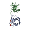
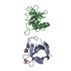
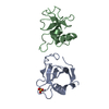
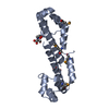
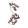
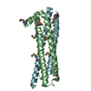
- PDB-5jyn: Structure of the transmembrane domain of HIV-1 gp41 in bicelle -

+
Open data


ID or keywords:

Loading...

-
Basic information

Entry
Database: PDB / ID: 5jyn
TitleStructure of the transmembrane domain of HIV-1 gp41 in bicelle
ComponentsEnvelope glycoprotein gp160
KeywordsVIRAL PROTEIN / transmembrane domain / lipid bilayer / transmembrane trimer
Function / homology
Function and homology information


host cell endosome membrane / clathrin-dependent endocytosis of virus by host cell / virus-mediated perturbation of host defense response / fusion of virus membrane with host endosome membrane / viral envelope / apoptotic process / host cell plasma membrane / structural molecule activity / virion membrane / plasma membrane
Similarity search - Function
Envelope glycoprotein Gp160 / Retroviral envelope protein / Retroviral envelope protein GP41-like / Gp120 core superfamily / Envelope glycoprotein GP120 / Human immunodeficiency virus 1, envelope glycoprotein Gp120
Similarity search - Domain/homology
Envelope glycoprotein gp160
Similarity search - Component
Biological speciesHuman immunodeficiency virus 1
MethodSOLUTION NMR / simulated annealing
AuthorsDev, J. / Fu, Q. / Park, D. / Chen, B. / Chou, J.J.
Funding support United States, China, 2items
OrganizationGrant numberCountry
National Institutes of Health/National Heart, Lung, and Blood Institute (NIH/NHLBI)HL103526 United States
CASXDB08030301 China
CitationJournal: Science / Year: 2016
Title: Structural basis for membrane anchoring of HIV-1 envelope spike.
Authors: Dev, J. / Park, D. / Fu, Q. / Chen, J. / Ha, H.J. / Ghantous, F. / Herrmann, T. / Chang, W. / Liu, Z. / Frey, G. / Seaman, M.S. / Chen, B. / Chou, J.J.
History
DepositionMay 14, 2016Deposition site: RCSB / Processing site: RCSB
Revision 1.0Jun 29, 2016Provider: repository / Type: Initial release
Revision 1.1Jul 6, 2016Group: Database references
Revision 1.2Jul 20, 2016Group: Database references
Revision 1.3Sep 20, 2017Group: Author supporting evidence / Database references / Structure summary
Category: citation / entity / pdbx_audit_support
Item: _citation.journal_id_CSD / _entity.pdbx_number_of_molecules / _pdbx_audit_support.funding_organization
Revision 1.4Dec 4, 2019Group: Author supporting evidence / Data collection / Category: pdbx_audit_support / pdbx_nmr_spectrometer
Item: _pdbx_audit_support.funding_organization / _pdbx_nmr_spectrometer.model
Revision 1.5Jun 14, 2023Group: Database references / Other / Category: database_2 / pdbx_database_status
Item: _database_2.pdbx_DOI / _database_2.pdbx_database_accession / _pdbx_database_status.status_code_nmr_data
Revision 1.6May 15, 2024Group: Data collection / Database references / Category: chem_comp_atom / chem_comp_bond / database_2 / Item: _database_2.pdbx_DOI

-
Structure visualization

Structure viewerMolecule:
MolmilJmol/JSmol

Downloads & links

-
Assembly

Deposited unit
A: Envelope glycoprotein gp160
B: Envelope glycoprotein gp160
C: Envelope glycoprotein gp160


Theoretical massNumber of molelcules
Total (without water)13,9523
Polymers13,9523
Non-polymers00
Water00
1


  • Idetical with deposited unit
  • defined by author
TypeNameSymmetry operationNumber
identity operation1_5551
Buried area3740 Å2
ΔGint-31 kcal/mol
Surface area9870 Å2
NMR ensembles
DataCriteria
Number of conformers (submitted / calculated)15 / 150structures with the lowest energy
RepresentativeModel #1lowest energy

-
Components

#1: Protein/peptide Envelope glycoprotein gp160


Mass: 4650.604 Da / Num. of mol.: 3 / Fragment: GP41 domain residues 670-709
Source method: isolated from a genetically manipulated source
Source: (gene. exp.) Human immunodeficiency virus 1 / Gene: env / Production host: Escherichia coli (E. coli) / Strain (production host): BL21 (DE3) / References: UniProt: Q74849

-
Experimental details

-
Experiment

ExperimentMethod: SOLUTION NMR
NMR experiment
Conditions-IDExperiment-IDSolution-IDSample stateSpectrometer-IDType
111isotropic13D TROSY HNCA
121isotropic13D TROSY HN(CO)CA
131isotropic13D TROSY HN(CA)CO
141isotropic13D TROSY HNCO
152isotropic33D 15N NOESY-TROSY-HSQC
162isotropic33D 13C NOESY-HSQC (Methyls)
173isotropic23D 15N NOESY-TROSY-HSQC (long mixing time)
181isotropic12D 15N TROSY-HSQC for GdDOTA titration
194isotropic13D 15N NOESY-TROSY-HSQC (short mixing time)

-
Sample preparation

Details
TypeSolution-IDContentsDetailsLabelSolvent system
bicelle10.8 mM [U-13C; U-15N; 85%-2H] HIV-1 gp41 Transmembrane Domain, 60 mM regular 1,2-Dimyristoyl-sn-Glycero-3-Phosphocholine (DMPC), 120 mM regular 1,2-Dihexanoyl-sn-Glycero-3-Phosphocholine (DHPC), 25 mM regular MES, 0.1 % regular NaN3, 95% H2O/5% D2OFor assignment of protein backbone resonancestriple labeled sample95% H2O/5% D2O
bicelle20.8 mM [U-13C; U-15N] HIV-1 gp41 Transmembrane Domain, 60 mM acyl chains 2H labeled 1,2-Dimyristoyl-sn-Glycero-3-Phosphocholine (DMPC), 120 mM acyl chains 2H labeled 1,2-Dihexanoyl-sn-Glycero-3-Phosphocholine (DHPC), 25 mM regular MES, 0.1 % regular NaN3, 95% H2O/5% D2OFor recording 3D 15N-edited and 13C-edited NOESYsdouble labeled sample95% H2O/5% D2O
bicelle30.4 mM [U-15N; U-2H] HIV-1 gp41 Transmembrane Domain, 0.4 mM [15% 13C] HIV-1 gp41 Transmembrane Domain, 60 mM acyl chains 2H labeled 1,2-Dimyristoyl-sn-Glycero-3-Phosphocholine (DMPC), 120 mM acyl chains 2H labeled 1,2-Dihexanoyl-sn-Glycero-3-Phosphocholine (DHPC), 25 mM regular MES, 0.1 % regular NaN3, 95% H2O/5% D2OFor assigning inter-molecular NOEsmixed labeled sample95% H2O/5% D2O
bicelle40.8 mM [U-15N] HIV-1 gp41 Transmembrane Domain, 60 mM acyl chains 2H labeled 1,2-Dimyristoyl-sn-Glycero-3-Phosphocholine (DMPC), 120 mM acyl chains 2H labeled 1,2-Dihexanoyl-sn-Glycero-3-Phosphocholine (DHPC), 25 mM regular MES, 0.1 % regular NaN3, 95% H2O/5% D2OFor assigning Arg sidechains15N labeled sample95% H2O/5% D2O
Sample
Conc. (mg/ml)ComponentIsotopic labelingSolution-ID
0.8 mMHIV-1 gp41 Transmembrane Domain[U-13C; U-15N; 85%-2H]1
60 mM1,2-Dimyristoyl-sn-Glycero-3-Phosphocholine (DMPC)regular1
120 mM1,2-Dihexanoyl-sn-Glycero-3-Phosphocholine (DHPC)regular1
25 mMMESregular1
0.1 %NaN3regular1
0.8 mMHIV-1 gp41 Transmembrane Domain[U-13C; U-15N]2
60 mM1,2-Dimyristoyl-sn-Glycero-3-Phosphocholine (DMPC)acyl chains 2H labeled2
120 mM1,2-Dihexanoyl-sn-Glycero-3-Phosphocholine (DHPC)acyl chains 2H labeled2
25 mMMESregular2
0.1 %NaN3regular2
0.4 mMHIV-1 gp41 Transmembrane Domain-1[U-15N; U-2H]3
0.4 mMHIV-1 gp41 Transmembrane Domain-2[15% 13C]3
60 mM1,2-Dimyristoyl-sn-Glycero-3-Phosphocholine (DMPC)acyl chains 2H labeled3
120 mM1,2-Dihexanoyl-sn-Glycero-3-Phosphocholine (DHPC)acyl chains 2H labeled3
25 mMMESregular3
0.1 %NaN3regular3
0.8 mMHIV-1 gp41 Transmembrane Domain[U-15N]4
60 mM1,2-Dimyristoyl-sn-Glycero-3-Phosphocholine (DMPC)acyl chains 2H labeled4
120 mM1,2-Dihexanoyl-sn-Glycero-3-Phosphocholine (DHPC)acyl chains 2H labeled4
25 mMMESregular4
0.1 %NaN3regular4
Sample conditionsIonic strength: 50 mM / Ionic strength err: 10 / Label: ambient / pH: 6.7 / PH err: 0.1 / Pressure: 1 atm / Temperature: 303 K / Temperature err: 0.1

-
NMR measurement

NMR spectrometer
TypeManufacturerModelField strength (MHz)Spectrometer-IDDetails
Bruker AVANCE IIBrukerAVANCE II6001cryogenic probe
Bruker AVANCE IIBrukerAVANCE II7502cryogenic probe
Bruker AVANCE IIIBrukerAVANCE III9003cryogenic probe

-
Processing

NMR software
NameVersionDeveloperClassification
XPLOR-NIH2.42G. Marius Clore , Guillermo Bermejo, , John Kuszewski, Charles D. Schwieters, and Nico Tjandrastructure calculation
CcpNMRCCPNchemical shift assignment
NMRPipeDelaglio, Grzesiek, Vuister, Zhu, Pfeifer and Baxprocessing
XEASYBartels et al.data analysis
TALOSCornilescu, Delaglio and Baxdata analysis
XPLOR-NIH2.42G. Marius Clore , Guillermo Bermejo, , John Kuszewski, Charles D. Schwieters, and Nico Tjandrarefinement
RefinementMethod: simulated annealing / Software ordinal: 2
NMR representativeSelection criteria: lowest energy
NMR ensembleConformer selection criteria: structures with the lowest energy
Conformers calculated total number: 150 / Conformers submitted total number: 15

+
About Yorodumi

-
News

-
Feb 9, 2022. New format data for meta-information of EMDB entries

New format data for meta-information of EMDB entries

  • Version 3 of the EMDB header file is now the official format.
  • The previous official version 1.9 will be removed from the archive.

Related info.:EMDB header

External links:wwPDB to switch to version 3 of the EMDB data model

-
Aug 12, 2020. Covid-19 info

Covid-19 info

URL: https://pdbj.org/emnavi/covid19.php

New page: Covid-19 featured information page in EM Navigator.

Related info.:Covid-19 info / Mar 5, 2020. Novel coronavirus structure data

+
Mar 5, 2020. Novel coronavirus structure data

Novel coronavirus structure data

Related info.:Yorodumi Speices / Aug 12, 2020. Covid-19 info

External links:COVID-19 featured content - PDBj / Molecule of the Month (242):Coronavirus Proteases

+
Jan 31, 2019. EMDB accession codes are about to change! (news from PDBe EMDB page)

EMDB accession codes are about to change! (news from PDBe EMDB page)

  • The allocation of 4 digits for EMDB accession codes will soon come to an end. Whilst these codes will remain in use, new EMDB accession codes will include an additional digit and will expand incrementally as the available range of codes is exhausted. The current 4-digit format prefixed with “EMD-” (i.e. EMD-XXXX) will advance to a 5-digit format (i.e. EMD-XXXXX), and so on. It is currently estimated that the 4-digit codes will be depleted around Spring 2019, at which point the 5-digit format will come into force.
  • The EM Navigator/Yorodumi systems omit the EMD- prefix.

Related info.:Q: What is EMD? / ID/Accession-code notation in Yorodumi/EM Navigator

External links:EMDB Accession Codes are Changing Soon! / Contact to PDBj

+
Jul 12, 2017. Major update of PDB

Major update of PDB

  • wwPDB released updated PDB data conforming to the new PDBx/mmCIF dictionary.
  • This is a major update changing the version number from 4 to 5, and with Remediation, in which all the entries are updated.
  • In this update, many items about electron microscopy experimental information are reorganized (e.g. em_software).
  • Now, EM Navigator and Yorodumi are based on the updated data.

External links:wwPDB Remediation / Enriched Model Files Conforming to OneDep Data Standards Now Available in the PDB FTP Archive

-
Yorodumi

Thousand views of thousand structures

  • Yorodumi is a browser for structure data from EMDB, PDB, SASBDB, etc.
  • This page is also the successor to EM Navigator detail page, and also detail information page/front-end page for Omokage search.
  • The word "yorodu" (or yorozu) is an old Japanese word meaning "ten thousand". "mi" (miru) is to see.

Related info.:EMDB / PDB / SASBDB / Comparison of 3 databanks / Yorodumi Search / Aug 31, 2016. New EM Navigator & Yorodumi / Yorodumi Papers / Jmol/JSmol / Function and homology information / Changes in new EM Navigator and Yorodumi

Read more