 Movie
Movie Controller


+ Open data

- Basic information
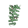
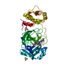
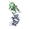
| Entry | Database: PDB / ID: 5jo8 | |||||||||
|---|---|---|---|---|---|---|---|---|---|---|
| Title | CEP104 TOG domain | |||||||||
 Components Components | CEP104 | |||||||||
 Keywords Keywords | STRUCTURAL PROTEIN / centriolar protein / TOG domain / CEP104 / microtubule binder | |||||||||
| Function / homology | TOG domain / TOG / Galactose-binding-like domain superfamily / Armadillo-like helical / Armadillo-type fold / TOG domain-containing protein Function and homology information Function and homology information | |||||||||
| Biological species |  | |||||||||
| Method |  X-RAY DIFFRACTION / SYNCHROTRON / SAD / Resolution: 1.4 Å X-RAY DIFFRACTION / SYNCHROTRON / SAD / Resolution: 1.4 Å | |||||||||
 Authors Authors | Rezabkova, L. / Kraatz, S.H.W. | |||||||||
| Funding support |  Belgium, Belgium,  Germany, 2items Germany, 2items
| |||||||||
 Citation Citation | Journal: J.Biol.Chem. / Year: 2016 Title: Biophysical and Structural Characterization of the Centriolar Protein Cep104 Interaction Network. Authors: Rezabkova, L. / Kraatz, S.H. / Akhmanova, A. / Steinmetz, M.O. / Kammerer, R.A. | |||||||||
| History |
|
- Structure visualization
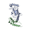
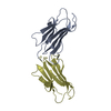
| Structure viewer | Molecule: MolmilJmol/JSmol |
|---|
- Downloads & links
-Download
| PDBx/mmCIF format | 5jo8.cif.gz | 70.3 KB | Display | PDBx/mmCIF format |
|---|---|---|---|---|
| PDB format | pdb5jo8.ent.gz | 50.6 KB | Display | PDB format |
| PDBx/mmJSON format | 5jo8.json.gz | Tree view | PDBx/mmJSON format | |
| Others |  Other downloads Other downloads |
-Validation report
| Arichive directory | https://data.pdbj.org/pub/pdb/validation_reports/jo/5jo8ftp://data.pdbj.org/pub/pdb/validation_reports/jo/5jo8 | HTTPS FTP |
|---|
-Related structure data
| Similar structure data |
|---|
-Links
 PDBj
PDBj- Assembly
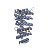
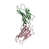
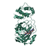
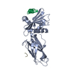
| Deposited unit | 
| |||||||||
|---|---|---|---|---|---|---|---|---|---|---|
| 1 |
| |||||||||
| Unit cell |
| |||||||||
| Components on special symmetry positions |
|
-Components
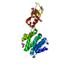
| #1: Protein | Mass: 31381.744 Da / Num. of mol.: 1 / Fragment: TOG domain, UNP residues 429-686 Source method: isolated from a genetically manipulated source Source: (gene. exp.)  |
|---|---|
| #2: Water | ChemComp-HOH /  Mass: 18.015 Da / Num. of mol.: 248 / Source method: isolated from a natural source / Formula: H2O Mass: 18.015 Da / Num. of mol.: 248 / Source method: isolated from a natural source / Formula: H2O |
-Experimental details
-Experiment
| Experiment | Method: X-RAY DIFFRACTION / Number of used crystals: 1 |
|---|
- Sample preparation
| Crystal | Density Matthews: 2.28 Å3/Da / Density % sol: 46 % |
|---|---|
| Crystal grow | Temperature: 293.15 K / Method: vapor diffusion, sitting drop / Details: 25% PEG 1500, 0.1 M MMT pH 5.0 |
-Data collection
| Diffraction | Mean temperature: 100 K | |||||||||
|---|---|---|---|---|---|---|---|---|---|---|
| Diffraction source | Source: SYNCHROTRON / Site: SLS  / Beamline: X06SA / Wavelength: 1.0, 0.96 / Beamline: X06SA / Wavelength: 1.0, 0.96 | |||||||||
| Detector | Type: DECTRIS PILATUS 2M-F / Detector: PIXEL / Date: Feb 28, 2015 | |||||||||
| Radiation | Protocol: SINGLE WAVELENGTH / Monochromatic (M) / Laue (L): M / Scattering type: x-ray | |||||||||
| Radiation wavelength |
| |||||||||
| Reflection | Resolution: 1.4→50 Å / Num. obs: 55668 / % possible obs: 97 % / Redundancy: 16.4 % / CC1/2: 1 / Rmerge(I) obs: 0.065 / Net I/σ(I): 19.02 | |||||||||
| Reflection shell | Resolution: 1.4→1.44 Å / Redundancy: 16.5 % / Rmerge(I) obs: 2.76 / Mean I/σ(I) obs: 1.05 / % possible all: 100 |
- Processing
| Software |
| |||||||||||||||||||||||||||||||||||||||||||||||||||||||||||||||||||||||||||||||||||||||||||||||||||||||||
|---|---|---|---|---|---|---|---|---|---|---|---|---|---|---|---|---|---|---|---|---|---|---|---|---|---|---|---|---|---|---|---|---|---|---|---|---|---|---|---|---|---|---|---|---|---|---|---|---|---|---|---|---|---|---|---|---|---|---|---|---|---|---|---|---|---|---|---|---|---|---|---|---|---|---|---|---|---|---|---|---|---|---|---|---|---|---|---|---|---|---|---|---|---|---|---|---|---|---|---|---|---|---|---|---|---|---|
| Refinement | Method to determine structure: SAD / Resolution: 1.4→43.772 Å / SU ML: 0.23 / Cross valid method: FREE R-VALUE / σ(F): 1.36 / Phase error: 29.12
| |||||||||||||||||||||||||||||||||||||||||||||||||||||||||||||||||||||||||||||||||||||||||||||||||||||||||
| Solvent computation | Shrinkage radii: 0.9 Å / VDW probe radii: 1.11 Å | |||||||||||||||||||||||||||||||||||||||||||||||||||||||||||||||||||||||||||||||||||||||||||||||||||||||||
| Refinement step | Cycle: LAST / Resolution: 1.4→43.772 Å
| |||||||||||||||||||||||||||||||||||||||||||||||||||||||||||||||||||||||||||||||||||||||||||||||||||||||||
| Refine LS restraints |
| |||||||||||||||||||||||||||||||||||||||||||||||||||||||||||||||||||||||||||||||||||||||||||||||||||||||||
| LS refinement shell |
|
