 Movie
Movie Controller

[English] 日本語
 Yorodumi
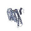
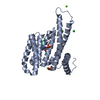
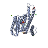
- PDB-5jip: Crystal structure of the Clostridium perfringens spore cortex lyt... -
+ Open data

- Basic information
| Entry | Database: PDB / ID: 5jip | ||||||
|---|---|---|---|---|---|---|---|
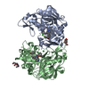
| Title | Crystal structure of the Clostridium perfringens spore cortex lytic enzyme SleM | ||||||
 Components Components | Cortical-lytic enzyme | ||||||
 Keywords Keywords | HYDROLASE / spore / cortex / peptidoglycan-lysin | ||||||
| Function / homology |  Function and homology information Function and homology informationcarbohydrate catabolic process / peptidoglycan catabolic process / cell wall macromolecule catabolic process / lysozyme / lysozyme activity / metal ion binding Similarity search - Function | ||||||
| Biological species |   Clostridium perfringens (bacteria) Clostridium perfringens (bacteria) | ||||||
| Method |  X-RAY DIFFRACTION / MOLECULAR REPLACEMENT / Resolution: 1.8 Å X-RAY DIFFRACTION / MOLECULAR REPLACEMENT / Resolution: 1.8 Å | ||||||
 Authors Authors | Chirgadze, D.Y. / Christie, G. / Ustok, F.I. / Al-Riyami, B. / Stott, K. | ||||||
 Citation Citation | Journal: Proteins / Year: 2016 Title: The crystal structure of Clostridium perfringens SleM, a muramidase involved in cortical hydrolysis during spore germination. Authors: Al-Riyami, B. / Ustok, F.I. / Stott, K. / Chirgadze, D.Y. / Christie, G. | ||||||
| History |
|
- Structure visualization
| Structure viewer | Molecule: MolmilJmol/JSmol |
|---|
- Downloads & links
-Download
| PDBx/mmCIF format | 5jip.cif.gz | 275.7 KB | Display | PDBx/mmCIF format |
|---|---|---|---|---|
| PDB format | pdb5jip.ent.gz | 222.7 KB | Display | PDB format |
| PDBx/mmJSON format | 5jip.json.gz | Tree view | PDBx/mmJSON format | |
| Others |  Other downloads Other downloads |
-Validation report
| Summary document | 5jip_validation.pdf.gz | 445.1 KB | Display | wwPDB validaton report |
|---|---|---|---|---|
| Full document | 5jip_full_validation.pdf.gz | 447.9 KB | Display | |
| Data in XML | 5jip_validation.xml.gz | 36.4 KB | Display | |
| Data in CIF | 5jip_validation.cif.gz | 58.5 KB | Display | |
| Arichive directory | https://data.pdbj.org/pub/pdb/validation_reports/ji/5jipftp://data.pdbj.org/pub/pdb/validation_reports/ji/5jip | HTTPS FTP |
-Related structure data
| Related structure data |  1jfxS S: Starting model for refinement |
|---|---|
| Similar structure data |


-Links
 PDBj
PDBj- Assembly
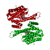
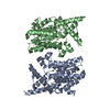
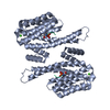
| Deposited unit | 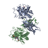
| ||||||||
|---|---|---|---|---|---|---|---|---|---|
| 1 |
| ||||||||
| Unit cell |
|
-Components
| #1: Protein | Mass: 37584.328 Da / Num. of mol.: 2 Source method: isolated from a genetically manipulated source Source: (gene. exp.) Clostridium perfringens (bacteria) / Gene: sleM / Plasmid: pBADcLIC-SleMDetails (production host): arabinose inducible expression plasmid compatible with ligation independent cloning Production host: #2: Chemical | ChemComp-MES /   Mass: 195.237 Da / Num. of mol.: 4 / Source method: obtained synthetically / Formula: C6H13NO4S / Comment: pH buffer*YM Mass: 195.237 Da / Num. of mol.: 4 / Source method: obtained synthetically / Formula: C6H13NO4S / Comment: pH buffer*YM#3: Chemical |   Mass: 24.305 Da / Num. of mol.: 2 / Source method: obtained synthetically / Formula: Mg Mass: 24.305 Da / Num. of mol.: 2 / Source method: obtained synthetically / Formula: Mg#4: Water | ChemComp-HOH / |  Mass: 18.015 Da / Num. of mol.: 1139 / Source method: isolated from a natural source / Formula: H2O Mass: 18.015 Da / Num. of mol.: 1139 / Source method: isolated from a natural source / Formula: H2O |
|---|
-Experimental details
-Experiment
| Experiment | Method: X-RAY DIFFRACTION / Number of used crystals: 1 |
|---|
- Sample preparation
| Crystal | Density Matthews: 2.44 Å3/Da / Density % sol: 49.7 % |
|---|---|
| Crystal grow | Temperature: 292 K / Method: vapor diffusion, sitting drop / pH: 6 Details: 0.1 M MES buffer, pH 6.0, 0.25 M magnesium chloride, 16% (w/v) PEG6000 |
-Data collection
| Diffraction | Mean temperature: 100 K |
|---|---|
| Diffraction source | Source: ROTATING ANODE / Type: BRUKER AXS MICROSTAR / Wavelength: 1.5418 Å |
| Detector | Type: Bruker Platinum 135 / Detector: CCD / Date: Dec 5, 2014 / Details: HELIOS MX |
| Radiation | Protocol: SINGLE WAVELENGTH / Monochromatic (M) / Laue (L): M / Scattering type: x-ray |
| Radiation wavelength | Wavelength: 1.5418 Å / Relative weight: 1 |
| Reflection | Resolution: 1.8→42.4 Å / Num. obs: 66317 / % possible obs: 99.6 % / Redundancy: 3.9 % / Biso Wilson estimate: 14.1 Å2 / Rsym value: 0.074 / Net I/σ(I): 24.5 |
| Reflection shell | Resolution: 1.8→1.9 Å / Redundancy: 2.9 % / Rmerge(I) obs: 0.357 / Mean I/σ(I) obs: 5 / % possible all: 98.2 |
- Processing
| Software |
| |||||||||||||||||||||||||||||||||||||||||||||||||||||||||||||||||||||||||||||||||||||||||||||||||||||||||||||||||||||||||||||||||||||||||||||||||||||||||||||||||||||||||||||||
|---|---|---|---|---|---|---|---|---|---|---|---|---|---|---|---|---|---|---|---|---|---|---|---|---|---|---|---|---|---|---|---|---|---|---|---|---|---|---|---|---|---|---|---|---|---|---|---|---|---|---|---|---|---|---|---|---|---|---|---|---|---|---|---|---|---|---|---|---|---|---|---|---|---|---|---|---|---|---|---|---|---|---|---|---|---|---|---|---|---|---|---|---|---|---|---|---|---|---|---|---|---|---|---|---|---|---|---|---|---|---|---|---|---|---|---|---|---|---|---|---|---|---|---|---|---|---|---|---|---|---|---|---|---|---|---|---|---|---|---|---|---|---|---|---|---|---|---|---|---|---|---|---|---|---|---|---|---|---|---|---|---|---|---|---|---|---|---|---|---|---|---|---|---|---|---|---|
| Refinement | Method to determine structure: MOLECULAR REPLACEMENT Starting model: 1JFX Resolution: 1.8→42.397 Å / SU ML: 0.18 / Cross valid method: FREE R-VALUE / σ(F): 1.33 / Phase error: 18.01
| |||||||||||||||||||||||||||||||||||||||||||||||||||||||||||||||||||||||||||||||||||||||||||||||||||||||||||||||||||||||||||||||||||||||||||||||||||||||||||||||||||||||||||||||
| Solvent computation | Shrinkage radii: 0.9 Å / VDW probe radii: 1.11 Å | |||||||||||||||||||||||||||||||||||||||||||||||||||||||||||||||||||||||||||||||||||||||||||||||||||||||||||||||||||||||||||||||||||||||||||||||||||||||||||||||||||||||||||||||
| Refinement step | Cycle: LAST / Resolution: 1.8→42.397 Å
| |||||||||||||||||||||||||||||||||||||||||||||||||||||||||||||||||||||||||||||||||||||||||||||||||||||||||||||||||||||||||||||||||||||||||||||||||||||||||||||||||||||||||||||||
| Refine LS restraints |
| |||||||||||||||||||||||||||||||||||||||||||||||||||||||||||||||||||||||||||||||||||||||||||||||||||||||||||||||||||||||||||||||||||||||||||||||||||||||||||||||||||||||||||||||
| LS refinement shell |
|
