[English] 日本語
Yorodumi
- PDB-5jht: Apo form of influenza strain H1N1 polymerase acidic subunit N-ter... -

+
Open data


ID or keywords:

Loading...

-
Basic information

Entry
Database: PDB / ID: 5jht
Title
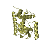
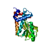
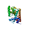
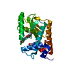
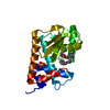
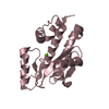
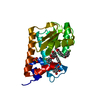
Apo form of influenza strain H1N1 polymerase acidic subunit N-terminal region crystallized with potassium sodium tartrate
ComponentsPolymerase acidic protein
KeywordsHYDROLASE
Function / homology
Function and homology information


cRNA Synthesis / Influenza Infection / Fusion of the Influenza Virion to the Host Cell Endosome / Release / Budding / Packaging of Eight RNA Segments / Uncoating of the Influenza Virion / Entry of Influenza Virion into Host Cell via Endocytosis / Viral RNP Complexes in the Host Cell Nucleus / vRNA Synthesis ...cRNA Synthesis / Influenza Infection / Fusion of the Influenza Virion to the Host Cell Endosome / Release / Budding / Packaging of Eight RNA Segments / Uncoating of the Influenza Virion / Entry of Influenza Virion into Host Cell via Endocytosis / Viral RNP Complexes in the Host Cell Nucleus / vRNA Synthesis / Transport of Ribonucleoproteins into the Host Nucleus / NEP/NS2 Interacts with the Cellular Export Machinery / vRNP Assembly / Viral Messenger RNA Synthesis / cap snatching / symbiont-mediated suppression of host mRNA transcription via inhibition of RNA polymerase II activity / Viral mRNA Translation / endonuclease activity / host cell cytoplasm / Hydrolases; Acting on ester bonds / viral RNA genome replication / DNA-templated transcription / host cell nucleus / RNA binding / extracellular region / metal ion binding
Similarity search - Function
Influenza RNA-dependent RNA polymerase subunit PA, endonuclease domain / Restriction Endonuclease / Polymerase acidic protein / Influenza RNA-dependent RNA polymerase subunit PA / Influenza RNA-dependent RNA polymerase subunit PA, endonuclease domain / Influenza RNA-dependent RNA polymerase subunit PA / 3-Layer(aba) Sandwich / Alpha Beta
Similarity search - Domain/homology
: / L(+)-TARTARIC ACID / Polymerase acidic protein
Similarity search - Component
Biological speciesInfluenza A virus
MethodX-RAY DIFFRACTION / SYNCHROTRON / MOLECULAR REPLACEMENT / molecular replacement / Resolution: 1.751 Å
Model detailsRNA binding protein
AuthorsFudo, S. / Yamamoto, N. / Nukaga, M. / Odagiri, T. / Tashiro, M. / Hoshino, T.
Funding support Japan, 1items
OrganizationGrant numberCountry
Japan Society for the Promotion of Science24590548 Japan
CitationJournal: Cryst.Growth Des. / Year: 2017
Title: Influence of Precipitants on Molecular Arrangements and Space Groups of Protein Crystals
Authors: Fudo, S. / Fei, Q. / Nukaga, M. / Hoshino, T.
History
DepositionApr 21, 2016Deposition site: RCSB / Processing site: PDBJ
Revision 1.0May 11, 2016Provider: repository / Type: Initial release
Revision 1.1Mar 1, 2017Group: Database references
Revision 1.2Feb 19, 2020Group: Data collection / Category: diffrn_source / Item: _diffrn_source.pdbx_synchrotron_site
Revision 1.3Nov 8, 2023Group: Data collection / Database references ...Data collection / Database references / Derived calculations / Refinement description
Category: chem_comp_atom / chem_comp_bond ...chem_comp_atom / chem_comp_bond / database_2 / pdbx_initial_refinement_model / pdbx_struct_conn_angle / struct_conn
Item: _database_2.pdbx_DOI / _database_2.pdbx_database_accession ..._database_2.pdbx_DOI / _database_2.pdbx_database_accession / _pdbx_struct_conn_angle.ptnr1_auth_comp_id / _pdbx_struct_conn_angle.ptnr1_auth_seq_id / _pdbx_struct_conn_angle.ptnr1_label_asym_id / _pdbx_struct_conn_angle.ptnr1_label_atom_id / _pdbx_struct_conn_angle.ptnr1_label_comp_id / _pdbx_struct_conn_angle.ptnr3_auth_comp_id / _pdbx_struct_conn_angle.ptnr3_auth_seq_id / _pdbx_struct_conn_angle.ptnr3_label_asym_id / _pdbx_struct_conn_angle.ptnr3_label_atom_id / _pdbx_struct_conn_angle.ptnr3_label_comp_id / _pdbx_struct_conn_angle.value / _struct_conn.pdbx_dist_value / _struct_conn.ptnr1_label_atom_id / _struct_conn.ptnr2_auth_comp_id / _struct_conn.ptnr2_auth_seq_id / _struct_conn.ptnr2_label_asym_id / _struct_conn.ptnr2_label_atom_id / _struct_conn.ptnr2_label_comp_id

-
Structure visualization

Structure viewerMolecule:
MolmilJmol/JSmol

Downloads & links

-
Assembly

Deposited unit
A: Polymerase acidic protein
hetero molecules


Theoretical massNumber of molelcules
Total (without water)22,5604
Polymers22,3001
Non-polymers2603
Water3,549197
1


  • Idetical with deposited unit
  • defined by author
TypeNameSymmetry operationNumber
identity operation1_555x,y,z1
Unit cell
Length a, b, c (Å)66.859, 66.859, 128.003
Angle α, β, γ (deg.)90.000, 90.000, 90.000
Int Tables number92
Space group name H-MP41212

-
Components

#1: Protein Polymerase acidic protein / RNA-directed RNA polymerase subunit P2


Mass: 22300.494 Da / Num. of mol.: 1 / Fragment: UNP RESIDUES 1-50, 73-197
Source method: isolated from a genetically manipulated source
Source: (gene. exp.) Influenza A virus / Strain: A/Puerto Rico/8/1934 H1N1 / Gene: PA / Production host: Escherichia coli (E. coli) / References: UniProt: P03433
#2: Chemical ChemComp-MN / MANGANESE (II) ION


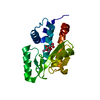
Mass: 54.938 Da / Num. of mol.: 2 / Source method: obtained synthetically / Formula: Mn
#3: Chemical ChemComp-TLA / L(+)-TARTARIC ACID


Mass: 150.087 Da / Num. of mol.: 1 / Source method: obtained synthetically / Formula: C4H6O6
#4: Water ChemComp-HOH / water


Mass: 18.015 Da / Num. of mol.: 197 / Source method: isolated from a natural source / Formula: H2O

-
Experimental details

-
Experiment

ExperimentMethod: X-RAY DIFFRACTION / Number of used crystals: 1

-
Sample preparation

CrystalDensity Matthews: 3.21 Å3/Da / Density % sol: 61.65 %
Crystal growTemperature: 291 K / Method: vapor diffusion, hanging drop / pH: 6 / Details: 0.1 M MES pH 6.0, 0.8 M Potassium sodium tartrate

-
Data collection

DiffractionMean temperature: 100 K
Diffraction sourceSource: SYNCHROTRON / Site: Photon Factory / Beamline: BL-5A / Wavelength: 1 Å
DetectorType: ADSC QUANTUM 315r / Detector: CCD / Date: Nov 19, 2015
RadiationMonochromator: Numerical link type Si(111) double crystal monochromator, direct water cooling
Protocol: SINGLE WAVELENGTH / Monochromatic (M) / Laue (L): M / Scattering type: x-ray
Radiation wavelengthWavelength: 1 Å / Relative weight: 1
ReflectionResolution: 1.75→50 Å / Num. obs: 29953 / % possible obs: 99.6 % / Observed criterion σ(F): 0 / Observed criterion σ(I): -3 / Redundancy: 14 % / Rmerge(I) obs: 0.059 / Rsym value: 0.059 / Net I/σ(I): 44.219
Reflection shellResolution: 1.75→1.78 Å / Redundancy: 14.3 % / Rmerge(I) obs: 1.359 / Mean I/σ(I) obs: 2.34 / % possible all: 100

-
Phasing

PhasingMethod: molecular replacement
Phasing MR
Highest resolutionLowest resolution
Rotation32.35 Å2.12 Å

-
Processing

Software
NameVersionClassification
HKL-2000data collection
HKL-2000data scaling
MOLREP11.0.05phasing
PHENIX1.8.1_1168refinement
PDB_EXTRACT3.2data extraction
RefinementMethod to determine structure: MOLECULAR REPLACEMENT
Starting model: 4ZQQ
Resolution: 1.751→32.345 Å / SU ML: 0.2 / Cross valid method: FREE R-VALUE / σ(F): 1.34 / Phase error: 23.49
RfactorNum. reflection% reflection
Rfree0.2379 1519 5.08 %
Rwork0.1943 --
obs0.1965 29878 99.56 %
Solvent computationShrinkage radii: 0.9 Å / VDW probe radii: 1.11 Å
Displacement parametersBiso max: 93.24 Å2 / Biso mean: 35.97 Å2 / Biso min: 19 Å2
Refinement stepCycle: final / Resolution: 1.751→32.345 Å
ProteinNucleic acidLigandSolventTotal
Num. atoms1493 0 12 197 1702
Biso mean--37.69 45.84 -
Num. residues----181
Refine LS restraints
Refine-IDTypeDev idealNumber
X-RAY DIFFRACTIONf_bond_d0.0171532
X-RAY DIFFRACTIONf_angle_d1.6342056
X-RAY DIFFRACTIONf_chiral_restr0.123220
X-RAY DIFFRACTIONf_plane_restr0.008264
X-RAY DIFFRACTIONf_dihedral_angle_d14.58584
LS refinement shell

Refine-ID: X-RAY DIFFRACTION / Total num. of bins used: 11

Resolution (Å)Rfactor RfreeNum. reflection RfreeRfactor RworkNum. reflection RworkNum. reflection all% reflection obs (%)
1.7513-1.80780.3131340.257225472681100
1.8078-1.87240.26881360.229225182654100
1.8724-1.94740.29481340.226625362670100
1.9474-2.0360.25621290.218725782707100
2.036-2.14330.251430.203325202663100
2.1433-2.27760.24571490.197925502699100
2.2776-2.45340.24041400.198225732713100
2.4534-2.70020.24891370.201425892726100
2.7002-3.09060.23741410.190225992740100
3.0906-3.89280.21841420.17932634277699
3.8928-32.35020.23181340.18982715284996

+
About Yorodumi

-
News

-
Feb 9, 2022. New format data for meta-information of EMDB entries

New format data for meta-information of EMDB entries

  • Version 3 of the EMDB header file is now the official format.
  • The previous official version 1.9 will be removed from the archive.

Related info.:EMDB header

External links:wwPDB to switch to version 3 of the EMDB data model

-
Aug 12, 2020. Covid-19 info

Covid-19 info

URL: https://pdbjlvh1.pdbj.org/emnavi/covid19.php

New page: Covid-19 featured information page in EM Navigator.

Related info.:Covid-19 info / Mar 5, 2020. Novel coronavirus structure data

+
Mar 5, 2020. Novel coronavirus structure data

Novel coronavirus structure data

Related info.:Yorodumi Speices / Aug 12, 2020. Covid-19 info

External links:COVID-19 featured content - PDBj / Molecule of the Month (242):Coronavirus Proteases

+
Jan 31, 2019. EMDB accession codes are about to change! (news from PDBe EMDB page)

EMDB accession codes are about to change! (news from PDBe EMDB page)

  • The allocation of 4 digits for EMDB accession codes will soon come to an end. Whilst these codes will remain in use, new EMDB accession codes will include an additional digit and will expand incrementally as the available range of codes is exhausted. The current 4-digit format prefixed with “EMD-” (i.e. EMD-XXXX) will advance to a 5-digit format (i.e. EMD-XXXXX), and so on. It is currently estimated that the 4-digit codes will be depleted around Spring 2019, at which point the 5-digit format will come into force.
  • The EM Navigator/Yorodumi systems omit the EMD- prefix.

Related info.:Q: What is EMD? / ID/Accession-code notation in Yorodumi/EM Navigator

External links:EMDB Accession Codes are Changing Soon! / Contact to PDBj

+
Jul 12, 2017. Major update of PDB

Major update of PDB

  • wwPDB released updated PDB data conforming to the new PDBx/mmCIF dictionary.
  • This is a major update changing the version number from 4 to 5, and with Remediation, in which all the entries are updated.
  • In this update, many items about electron microscopy experimental information are reorganized (e.g. em_software).
  • Now, EM Navigator and Yorodumi are based on the updated data.

External links:wwPDB Remediation / Enriched Model Files Conforming to OneDep Data Standards Now Available in the PDB FTP Archive

-
Yorodumi

Thousand views of thousand structures

  • Yorodumi is a browser for structure data from EMDB, PDB, SASBDB, etc.
  • This page is also the successor to EM Navigator detail page, and also detail information page/front-end page for Omokage search.
  • The word "yorodu" (or yorozu) is an old Japanese word meaning "ten thousand". "mi" (miru) is to see.

Related info.:EMDB / PDB / SASBDB / Comparison of 3 databanks / Yorodumi Search / Aug 31, 2016. New EM Navigator & Yorodumi / Yorodumi Papers / Jmol/JSmol / Function and homology information / Changes in new EM Navigator and Yorodumi

Read more