 Movie
Movie Controller

[English] 日本語
 Yorodumi
- PDB-5jfm: Crystal structure of Rhodopseudomonas palustris propionaldehyde d... -
+ Open data

- Basic information
| Entry | Database: PDB / ID: 5jfm | ||||||
|---|---|---|---|---|---|---|---|
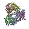
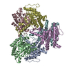
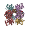
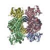
| Title | Crystal structure of Rhodopseudomonas palustris propionaldehyde dehydrogenase with bound propionyl-CoA | ||||||
 Components Components | Aldehyde dehydrogenase | ||||||
 Keywords Keywords | OXIDOREDUCTASE / acylating aldehyde dehydrogenase / propionylcysteine / bacterial microcompartments | ||||||
| Function / homology |  Function and homology information Function and homology information | ||||||
| Biological species |  Rhodopseudomonas palustris (phototrophic) Rhodopseudomonas palustris (phototrophic) | ||||||
| Method |  X-RAY DIFFRACTION / SYNCHROTRON / MOLECULAR REPLACEMENT / Resolution: 2.516 Å X-RAY DIFFRACTION / SYNCHROTRON / MOLECULAR REPLACEMENT / Resolution: 2.516 Å | ||||||
 Authors Authors | Zarzycki, J. / Sutter, M. / Kerfeld, C.A. | ||||||
 Citation Citation | Journal: Sci Rep / Year: 2017 Title: In Vitro Characterization and Concerted Function of Three Core Enzymes of a Glycyl Radical Enzyme - Associated Bacterial Microcompartment. Authors: Zarzycki, J. / Sutter, M. / Cortina, N.S. / Erb, T.J. / Kerfeld, C.A. | ||||||
| History |
|
- Structure visualization
| Structure viewer | Molecule: MolmilJmol/JSmol |
|---|
- Downloads & links
-Download
| PDBx/mmCIF format | 5jfm.cif.gz | 684.5 KB | Display | PDBx/mmCIF format |
|---|---|---|---|---|
| PDB format | pdb5jfm.ent.gz | 564 KB | Display | PDB format |
| PDBx/mmJSON format | 5jfm.json.gz | Tree view | PDBx/mmJSON format | |
| Others |  Other downloads Other downloads |
-Validation report
| Arichive directory | https://data.pdbj.org/pub/pdb/validation_reports/jf/5jfmftp://data.pdbj.org/pub/pdb/validation_reports/jf/5jfm | HTTPS FTP |
|---|
-Related structure data
| Related structure data |  5jflC  5jfnC  4c3sS C: citing same article ( S: Starting model for refinement |
|---|---|
| Similar structure data |


-Links
 PDBj
PDBj


- Assembly
| Deposited unit | 
| ||||||||
|---|---|---|---|---|---|---|---|---|---|
| 1 | 
| ||||||||
| 2 | 
| ||||||||
| Unit cell |
|
-Components
| #1: Protein | Mass: 55523.531 Da / Num. of mol.: 8 Source method: isolated from a genetically manipulated source Source: (gene. exp.) Rhodopseudomonas palustris (strain BisB18) (phototrophic)Strain: BisB18 / Gene: RPC_1174 / Production host: #2: Chemical | ChemComp-1VU /   Mass: 823.597 Da / Num. of mol.: 7 / Source method: isolated from a natural source / Formula: C24H40N7O17P3S Mass: 823.597 Da / Num. of mol.: 7 / Source method: isolated from a natural source / Formula: C24H40N7O17P3S#3: Chemical | ChemComp-COA / |   Mass: 767.534 Da / Num. of mol.: 1 / Source method: obtained synthetically / Formula: C21H36N7O16P3S Mass: 767.534 Da / Num. of mol.: 1 / Source method: obtained synthetically / Formula: C21H36N7O16P3S#4: Water | ChemComp-HOH / |  Mass: 18.015 Da / Num. of mol.: 1091 / Source method: isolated from a natural source / Formula: H2O Mass: 18.015 Da / Num. of mol.: 1091 / Source method: isolated from a natural source / Formula: H2O |
|---|
-Experimental details
-Experiment
| Experiment | Method: X-RAY DIFFRACTION / Number of used crystals: 1 |
|---|
- Sample preparation
| Crystal | Density Matthews: 2.39 Å3/Da / Density % sol: 48.6 % |
|---|---|
| Crystal grow | Temperature: 295 K / Method: vapor diffusion, sitting drop / pH: 4.8 Details: 50 mM sodium citrate, pH 4.8, 4% w/v PEG8000, 5 mM propionyl-CoA |
-Data collection
| Diffraction | Mean temperature: 100 K |
|---|---|
| Diffraction source | Source: SYNCHROTRON / Site: ALS  / Beamline: 5.0.2 / Wavelength: 1.00001 Å / Beamline: 5.0.2 / Wavelength: 1.00001 Å |
| Detector | Type: ADSC QUANTUM 315r / Detector: CCD / Date: Dec 20, 2014 |
| Radiation | Protocol: SINGLE WAVELENGTH / Monochromatic (M) / Laue (L): M / Scattering type: x-ray |
| Radiation wavelength | Wavelength: 1.00001 Å / Relative weight: 1 |
| Reflection | Resolution: 2.516→39.26 Å / Num. obs: 135429 / % possible obs: 95.5 % / Redundancy: 2.3 % / Rmerge(I) obs: 0.072 / Net I/σ(I): 11.3 |
| Reflection shell | Resolution: 2.52→2.65 Å / Redundancy: 2.2 % / Rmerge(I) obs: 0.502 / Mean I/σ(I) obs: 2.3 / % possible all: 91.8 |
- Processing
| Software |
| |||||||||||||||||||||||||||||||||||||||||||||||||||||||||||||||||||||||||||||||||||||||||||||||||||||||||
|---|---|---|---|---|---|---|---|---|---|---|---|---|---|---|---|---|---|---|---|---|---|---|---|---|---|---|---|---|---|---|---|---|---|---|---|---|---|---|---|---|---|---|---|---|---|---|---|---|---|---|---|---|---|---|---|---|---|---|---|---|---|---|---|---|---|---|---|---|---|---|---|---|---|---|---|---|---|---|---|---|---|---|---|---|---|---|---|---|---|---|---|---|---|---|---|---|---|---|---|---|---|---|---|---|---|---|
| Refinement | Method to determine structure: MOLECULAR REPLACEMENT Starting model: 4C3S Resolution: 2.516→39.259 Å / SU ML: 0.29 / Cross valid method: FREE R-VALUE / σ(F): 1.96 / Phase error: 23.25
| |||||||||||||||||||||||||||||||||||||||||||||||||||||||||||||||||||||||||||||||||||||||||||||||||||||||||
| Solvent computation | Shrinkage radii: 0.9 Å / VDW probe radii: 1.11 Å | |||||||||||||||||||||||||||||||||||||||||||||||||||||||||||||||||||||||||||||||||||||||||||||||||||||||||
| Refinement step | Cycle: LAST / Resolution: 2.516→39.259 Å
| |||||||||||||||||||||||||||||||||||||||||||||||||||||||||||||||||||||||||||||||||||||||||||||||||||||||||
| Refine LS restraints |
| |||||||||||||||||||||||||||||||||||||||||||||||||||||||||||||||||||||||||||||||||||||||||||||||||||||||||
| LS refinement shell |
|
