 Movie
Movie Controller

[English] 日本語
 Yorodumi
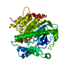
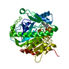
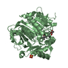
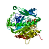
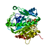
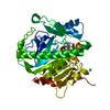
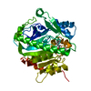
- PDB-5jad: Compound binding to Human Lipoprotein-Associated Phospholipase A2... -
+ Open data

- Basic information
| Entry | Database: PDB / ID: 5jad | ||||||
|---|---|---|---|---|---|---|---|
| Title | Compound binding to Human Lipoprotein-Associated Phospholipase A2 (Lp-PLA2)discovered through fragment screening | ||||||
 Components Components | Platelet-activating factor acetylhydrolase | ||||||
 Keywords Keywords | HYDROLASE / phospholipase / lipid metabolism | ||||||
| Function / homology |  Function and homology information Function and homology informationplasma lipoprotein particle oxidation / platelet activating factor catabolic process / : / 1-alkyl-2-acetylglycerophosphocholine esterase / 1-alkyl-2-acetylglycerophosphocholine esterase activity / platelet activating factor metabolic process / lipid oxidation / low-density lipoprotein particle / high-density lipoprotein particle / low-density lipoprotein particle remodeling ...plasma lipoprotein particle oxidation / platelet activating factor catabolic process / : / 1-alkyl-2-acetylglycerophosphocholine esterase / 1-alkyl-2-acetylglycerophosphocholine esterase activity / platelet activating factor metabolic process / lipid oxidation / low-density lipoprotein particle / high-density lipoprotein particle / low-density lipoprotein particle remodeling / phosphatidylcholine catabolic process / positive regulation of monocyte chemotaxis / peptide hormone processing / hydrolase activity, acting on ester bonds / Synthesis, secretion, and deacylation of Ghrelin / phospholipid binding / positive regulation of inflammatory response / extracellular region Similarity search - Function | ||||||
| Biological species |  Homo sapiens (human) Homo sapiens (human) | ||||||
| Method |  X-RAY DIFFRACTION / FOURIER SYNTHESIS / Resolution: 2.05 Å X-RAY DIFFRACTION / FOURIER SYNTHESIS / Resolution: 2.05 Å | ||||||
 Authors Authors | Day, P.J. / Woolford, A.J.-A. | ||||||
 Citation Citation | Journal: J.Med.Chem. / Year: 2016 Title: Exploitation of a Novel Binding Pocket in Human Lipoprotein-Associated Phospholipase A2 (Lp-PLA2) Discovered through X-ray Fragment Screening. Authors: Woolford, A.J. / Pero, J.E. / Aravapalli, S. / Berdini, V. / Coyle, J.E. / Day, P.J. / Dodson, A.M. / Grondin, P. / Holding, F.P. / Lee, L.Y. / Li, P. / Manas, E.S. / Marino, J. / Martin, A. ...Authors: Woolford, A.J. / Pero, J.E. / Aravapalli, S. / Berdini, V. / Coyle, J.E. / Day, P.J. / Dodson, A.M. / Grondin, P. / Holding, F.P. / Lee, L.Y. / Li, P. / Manas, E.S. / Marino, J. / Martin, A.C. / McCleland, B.W. / McMenamin, R.L. / Murray, C.W. / Neipp, C.E. / Page, L.W. / Patel, V.K. / Potvain, F. / Rich, S. / Rivero, R.A. / Smith, K. / Somers, D.O. / Trottet, L. / Velagaleti, R. / Williams, G. / Xie, R. | ||||||
| History |
|
- Structure visualization
| Structure viewer | Molecule: MolmilJmol/JSmol |
|---|
- Downloads & links
-Download
| PDBx/mmCIF format | 5jad.cif.gz | 169.3 KB | Display | PDBx/mmCIF format |
|---|---|---|---|---|
| PDB format | pdb5jad.ent.gz | 130.7 KB | Display | PDB format |
| PDBx/mmJSON format | 5jad.json.gz | Tree view | PDBx/mmJSON format | |
| Others |  Other downloads Other downloads |
-Validation report
| Arichive directory | https://data.pdbj.org/pub/pdb/validation_reports/ja/5jadftp://data.pdbj.org/pub/pdb/validation_reports/ja/5jad | HTTPS FTP |
|---|
-Related structure data
| Related structure data |  5jahC 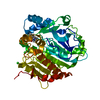 5jalC  5janC  5jaoC 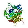 5japC 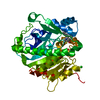 5jarC 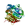 5jasC 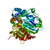 5jatC 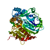 5jauC C: citing same article ( |
|---|---|
| Similar structure data |


-Links
 PDBj
PDBj


- Assembly
| Deposited unit | 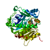
| ||||||||||||
|---|---|---|---|---|---|---|---|---|---|---|---|---|---|
| 1 |
| ||||||||||||
| Unit cell |
| ||||||||||||
| Components on special symmetry positions |
|
-Components
-Protein , 1 types, 1 molecules A
| #1: Protein | Mass: 44203.129 Da / Num. of mol.: 1 Source method: isolated from a genetically manipulated source Source: (gene. exp.) Homo sapiens (human) / Gene: PLA2G7, PAFAH / Production host:  References: UniProt: Q13093, 1-alkyl-2-acetylglycerophosphocholine esterase |
|---|
-Non-polymers , 5 types, 147 molecules 


| #2: Chemical | ChemComp-MG /  Mass: 24.305 Da / Num. of mol.: 1 / Source method: obtained synthetically / Formula: Mg Mass: 24.305 Da / Num. of mol.: 1 / Source method: obtained synthetically / Formula: Mg |
|---|---|
| #3: Chemical | ChemComp-CL /  Mass: 35.453 Da / Num. of mol.: 1 / Source method: obtained synthetically / Formula: Cl Mass: 35.453 Da / Num. of mol.: 1 / Source method: obtained synthetically / Formula: Cl |
| #4: Chemical | ChemComp-DMS /  Mass: 78.133 Da / Num. of mol.: 1 / Source method: obtained synthetically / Formula: C2H6OS / Comment: DMSO, precipitant*YM Mass: 78.133 Da / Num. of mol.: 1 / Source method: obtained synthetically / Formula: C2H6OS / Comment: DMSO, precipitant*YM |
| #5: Chemical | ChemComp-FB2 /  Mass: 157.190 Da / Num. of mol.: 1 / Source method: obtained synthetically / Formula: C6H7NO2S Mass: 157.190 Da / Num. of mol.: 1 / Source method: obtained synthetically / Formula: C6H7NO2S |
| #6: Water | ChemComp-HOH / Mass: 18.015 Da / Num. of mol.: 143 / Source method: isolated from a natural source / Formula: H2O |
-Experimental details
-Experiment
| Experiment | Method: X-RAY DIFFRACTION / Number of used crystals: 1 |
|---|
- Sample preparation
| Crystal | Density Matthews: 2.42 Å3/Da / Density % sol: 49.12 % |
|---|---|
| Crystal grow | Temperature: 297 K / Method: vapor diffusion, sitting drop / pH: 7.4 Details: 1.2M NaCl, 0.1M HEPES/NaOHpH=7.4, 28.8%w/v PEG 3350 PH range: 7.4 |
-Data collection
| Diffraction | Mean temperature: 100 K |
|---|---|
| Diffraction source | Source: ROTATING ANODE / Type: RIGAKU FR-X / Wavelength: 1.5417 Å |
| Detector | Type: RIGAKU SATURN 944 / Detector: CCD / Date: Apr 15, 2010 / Details: VariMax VHF Arc)Sec optic |
| Radiation | Monochromator: VariMax VHF Arc)Sec optic / Protocol: SINGLE WAVELENGTH / Monochromatic (M) / Laue (L): M / Scattering type: x-ray |
| Radiation wavelength | Wavelength: 1.5417 Å / Relative weight: 1 |
| Reflection | Resolution: 2.05→64 Å / Num. obs: 103331 / % possible obs: 97.4 % / Observed criterion σ(F): 0 / Observed criterion σ(I): 0 / Redundancy: 3.4 % / Biso Wilson estimate: 28.81 Å2 / Rmerge(I) obs: 0.093 / Net I/σ(I): 8.6 |
| Reflection shell | Resolution: 2.05→2.07 Å / Redundancy: 3.4 % / Rmerge(I) obs: 0.519 / Mean I/σ(I) obs: 2.3 / % possible all: 88.7 |
- Processing
| Software |
| ||||||||||||||||||||||||||||||||||||||||||||||||||||||||||||||||||||||||||||||||||||||||||||||||||||||||||||||||||
|---|---|---|---|---|---|---|---|---|---|---|---|---|---|---|---|---|---|---|---|---|---|---|---|---|---|---|---|---|---|---|---|---|---|---|---|---|---|---|---|---|---|---|---|---|---|---|---|---|---|---|---|---|---|---|---|---|---|---|---|---|---|---|---|---|---|---|---|---|---|---|---|---|---|---|---|---|---|---|---|---|---|---|---|---|---|---|---|---|---|---|---|---|---|---|---|---|---|---|---|---|---|---|---|---|---|---|---|---|---|---|---|---|---|---|---|
| Refinement | Method to determine structure: FOURIER SYNTHESIS / Resolution: 2.05→32.31 Å / Cor.coef. Fo:Fc: 0.91 / Cor.coef. Fo:Fc free: 0.869 / SU R Cruickshank DPI: 0.248 / Cross valid method: FREE R-VALUE / σ(F): 0 / SU R Blow DPI: 0.244 / SU Rfree Blow DPI: 0.212 / SU Rfree Cruickshank DPI: 0.215 Details: Buster refinement was interspersed with rounds of rebuilding with COOT
| ||||||||||||||||||||||||||||||||||||||||||||||||||||||||||||||||||||||||||||||||||||||||||||||||||||||||||||||||||
| Displacement parameters | Biso mean: 33.676 Å2
| ||||||||||||||||||||||||||||||||||||||||||||||||||||||||||||||||||||||||||||||||||||||||||||||||||||||||||||||||||
| Refine analyze | Luzzati coordinate error obs: 0.33 Å | ||||||||||||||||||||||||||||||||||||||||||||||||||||||||||||||||||||||||||||||||||||||||||||||||||||||||||||||||||
| Refinement step | Cycle: 1 / Resolution: 2.05→32.31 Å
| ||||||||||||||||||||||||||||||||||||||||||||||||||||||||||||||||||||||||||||||||||||||||||||||||||||||||||||||||||
| Refine LS restraints |
| ||||||||||||||||||||||||||||||||||||||||||||||||||||||||||||||||||||||||||||||||||||||||||||||||||||||||||||||||||
| LS refinement shell | Highest resolution: 2.05 Å / Rfactor Rfree error: 0
| ||||||||||||||||||||||||||||||||||||||||||||||||||||||||||||||||||||||||||||||||||||||||||||||||||||||||||||||||||
| Refinement TLS params. | Method: refined / Origin x: 30.0021 Å / Origin y: 14.7021 Å / Origin z: 0.8666 Å
| ||||||||||||||||||||||||||||||||||||||||||||||||||||||||||||||||||||||||||||||||||||||||||||||||||||||||||||||||||
| Refinement TLS group | Selection details: { A|55 - A|425 } |
