 Movie
Movie Controller

[English] 日本語
 Yorodumi
- PDB-5j90: Structure of Fjoh_4558, a chitin-binding SusD homolog from Flavob... -
+ Open data

- Basic information
| Entry | Database: PDB / ID: 5j90 | ||||||
|---|---|---|---|---|---|---|---|
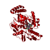
| Title | Structure of Fjoh_4558, a chitin-binding SusD homolog from Flavobacterium johnsoniae | ||||||
 Components Components | RagB/SusD domain protein | ||||||
 Keywords Keywords | Chitin-binding protein / Chitin-binding / SusD homolog / Bacteroidetes / Flavobacterium johnsoniae | ||||||
| Function / homology | SusD-like / Susd and RagB outer membrane lipoprotein / Serine Threonine Protein Phosphatase 5, Tetratricopeptide repeat - #390 / Serine Threonine Protein Phosphatase 5, Tetratricopeptide repeat / Alpha Horseshoe / Prokaryotic membrane lipoprotein lipid attachment site profile. / Tetratricopeptide-like helical domain superfamily / Mainly Alpha / RagB/SusD domain protein Function and homology information Function and homology information | ||||||
| Biological species |  Flavobacterium johnsoniae (bacteria) Flavobacterium johnsoniae (bacteria) | ||||||
| Method |  X-RAY DIFFRACTION / SYNCHROTRON / MOLECULAR REPLACEMENT / molecular replacement / Resolution: 1.3932 Å X-RAY DIFFRACTION / SYNCHROTRON / MOLECULAR REPLACEMENT / molecular replacement / Resolution: 1.3932 Å | ||||||
 Authors Authors | Koropatkin, N.M. | ||||||
 Citation Citation | Journal: Biotechnol Biofuels / Year: 2016 Title: A polysaccharide utilization locus from Flavobacterium johnsoniae enables conversion of recalcitrant chitin. Authors: Larsbrink, J. / Zhu, Y. / Kharade, S.S. / Kwiatkowski, K.J. / Eijsink, V.G. / Koropatkin, N.M. / McBride, M.J. / Pope, P.B. | ||||||
| History |
|
- Structure visualization
| Structure viewer | Molecule: MolmilJmol/JSmol |
|---|
- Downloads & links
-Download
| PDBx/mmCIF format | 5j90.cif.gz | 238.7 KB | Display | PDBx/mmCIF format |
|---|---|---|---|---|
| PDB format | pdb5j90.ent.gz | 185.4 KB | Display | PDB format |
| PDBx/mmJSON format | 5j90.json.gz | Tree view | PDBx/mmJSON format | |
| Others |  Other downloads Other downloads |
-Validation report
| Arichive directory | https://data.pdbj.org/pub/pdb/validation_reports/j9/5j90ftp://data.pdbj.org/pub/pdb/validation_reports/j9/5j90 | HTTPS FTP |
|---|
-Related structure data
| Related structure data | 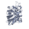 5j5uC 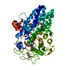 4f7aS C: citing same article ( S: Starting model for refinement |
|---|---|
| Similar structure data |


-Links
 PDBj
PDBj


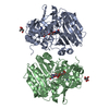
- Assembly
| Deposited unit | 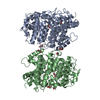
| ||||||||
|---|---|---|---|---|---|---|---|---|---|
| 1 |
| ||||||||
| Unit cell |
|
-Components
| #1: Protein | Mass: 56582.090 Da / Num. of mol.: 2 Source method: isolated from a genetically manipulated source Source: (gene. exp.) Flavobacterium johnsoniae (bacteria) / Strain: ATCC 17061 / DSM 2064 / UW101 / Gene: Fjoh_4558 / Plasmid: pETite N-his / Production host: #2: Chemical | ChemComp-EDO /   Mass: 62.068 Da / Num. of mol.: 11 / Source method: obtained synthetically / Formula: C2H6O2 Mass: 62.068 Da / Num. of mol.: 11 / Source method: obtained synthetically / Formula: C2H6O2#3: Water | ChemComp-HOH / |  Mass: 18.015 Da / Num. of mol.: 1514 / Source method: isolated from a natural source / Formula: H2O Mass: 18.015 Da / Num. of mol.: 1514 / Source method: isolated from a natural source / Formula: H2O |
|---|
-Experimental details
-Experiment
| Experiment | Method: X-RAY DIFFRACTION / Number of used crystals: 1 |
|---|
- Sample preparation
| Crystal | Density Matthews: 2.53 Å3/Da / Density % sol: 51.38 % |
|---|---|
| Crystal grow | Temperature: 293 K / Method: vapor diffusion, hanging drop / pH: 8.5 Details: 150 mM Tris-HCl pH 8.5, 27% sokolan CP5. These crystals were briefly soaked in 10mM acetylchitopentaose prior to freezing with a solution of 20% ethylene glycol, 80% crystallization media Temp details: room temp |
-Data collection
| Diffraction | Mean temperature: 100 K / Ambient temp details: nitrogen vapor | |||||||||||||||||||||||||||||||||||||||||||||||||||||||||||||||||||||||||||||||||||||||||||||||||||||||||
|---|---|---|---|---|---|---|---|---|---|---|---|---|---|---|---|---|---|---|---|---|---|---|---|---|---|---|---|---|---|---|---|---|---|---|---|---|---|---|---|---|---|---|---|---|---|---|---|---|---|---|---|---|---|---|---|---|---|---|---|---|---|---|---|---|---|---|---|---|---|---|---|---|---|---|---|---|---|---|---|---|---|---|---|---|---|---|---|---|---|---|---|---|---|---|---|---|---|---|---|---|---|---|---|---|---|---|
| Diffraction source | Source: SYNCHROTRON / Site: APS  / Beamline: 19-ID / Wavelength: 0.979 Å / Beamline: 19-ID / Wavelength: 0.979 Å | |||||||||||||||||||||||||||||||||||||||||||||||||||||||||||||||||||||||||||||||||||||||||||||||||||||||||
| Detector | Type: MARMOSAIC 225 mm CCD / Detector: CCD / Date: Jul 30, 2015 | |||||||||||||||||||||||||||||||||||||||||||||||||||||||||||||||||||||||||||||||||||||||||||||||||||||||||
| Radiation | Monochromator: 111 / Protocol: SINGLE WAVELENGTH / Monochromatic (M) / Laue (L): M / Scattering type: x-ray | |||||||||||||||||||||||||||||||||||||||||||||||||||||||||||||||||||||||||||||||||||||||||||||||||||||||||
| Radiation wavelength | Wavelength: 0.979 Å / Relative weight: 1 | |||||||||||||||||||||||||||||||||||||||||||||||||||||||||||||||||||||||||||||||||||||||||||||||||||||||||
| Reflection | Resolution: 1.39→50 Å / Num. obs: 215603 / % possible obs: 99.2 % / Redundancy: 7.9 % / Biso Wilson estimate: 11.53 Å2 / Rmerge(I) obs: 0.056 / Net I/av σ(I): 44.733 / Net I/σ(I): 12.4 | |||||||||||||||||||||||||||||||||||||||||||||||||||||||||||||||||||||||||||||||||||||||||||||||||||||||||
| Reflection shell |
|
-Phasing
| Phasing | Method: molecular replacement |
|---|
- Processing
| Software |
| |||||||||||||||||||||||||||||||||||||||||||||||||||||||||||||||||||||||||||||||||||||||||||||||||||||||||
|---|---|---|---|---|---|---|---|---|---|---|---|---|---|---|---|---|---|---|---|---|---|---|---|---|---|---|---|---|---|---|---|---|---|---|---|---|---|---|---|---|---|---|---|---|---|---|---|---|---|---|---|---|---|---|---|---|---|---|---|---|---|---|---|---|---|---|---|---|---|---|---|---|---|---|---|---|---|---|---|---|---|---|---|---|---|---|---|---|---|---|---|---|---|---|---|---|---|---|---|---|---|---|---|---|---|---|
| Refinement | Method to determine structure: MOLECULAR REPLACEMENT Starting model: 4F7A Resolution: 1.3932→36.574 Å / SU ML: 0.11 / Cross valid method: THROUGHOUT / σ(F): 1.34 / Phase error: 16.04
| |||||||||||||||||||||||||||||||||||||||||||||||||||||||||||||||||||||||||||||||||||||||||||||||||||||||||
| Solvent computation | Shrinkage radii: 0.9 Å / VDW probe radii: 1.11 Å | |||||||||||||||||||||||||||||||||||||||||||||||||||||||||||||||||||||||||||||||||||||||||||||||||||||||||
| Displacement parameters | Biso max: 56.9 Å2 / Biso mean: 15.7941 Å2 / Biso min: 5.75 Å2 | |||||||||||||||||||||||||||||||||||||||||||||||||||||||||||||||||||||||||||||||||||||||||||||||||||||||||
| Refinement step | Cycle: final / Resolution: 1.3932→36.574 Å
| |||||||||||||||||||||||||||||||||||||||||||||||||||||||||||||||||||||||||||||||||||||||||||||||||||||||||
| Refine LS restraints |
| |||||||||||||||||||||||||||||||||||||||||||||||||||||||||||||||||||||||||||||||||||||||||||||||||||||||||
| LS refinement shell | Refine-ID: X-RAY DIFFRACTION / Total num. of bins used: 14
|
