 Movie
Movie Controller

[English] 日本語
 Yorodumi
- PDB-5j68: Structure of Astrotactin-2, a conserved vertebrate-specific and p... -
+ Open data

- Basic information
| Entry | Database: PDB / ID: 5j68 | |||||||||
|---|---|---|---|---|---|---|---|---|---|---|
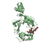
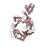
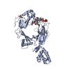
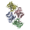
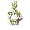
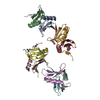
| Title | Structure of Astrotactin-2, a conserved vertebrate-specific and perforin-like membrane protein involved in neuronal development | |||||||||
 Components Components | Astrotactin-2 | |||||||||
 Keywords Keywords | MEMBRANE PROTEIN / MACPF / annexin-like / fibronectin / neural guidance | |||||||||
| Function / homology |  Function and homology information Function and homology informationinositol 1,3,4,5 tetrakisphosphate binding / cell pole / neuron cell-cell adhesion / clathrin-coated vesicle / neuron migration / late endosome / protein transport / cell cortex / perikaryon / early endosome ...inositol 1,3,4,5 tetrakisphosphate binding / cell pole / neuron cell-cell adhesion / clathrin-coated vesicle / neuron migration / late endosome / protein transport / cell cortex / perikaryon / early endosome / endosome / calcium ion binding / membrane Similarity search - Function | |||||||||
| Biological species |  Homo sapiens (human) Homo sapiens (human) | |||||||||
| Method |  X-RAY DIFFRACTION / SYNCHROTRON / MOLECULAR REPLACEMENT / Resolution: 5.221 Å X-RAY DIFFRACTION / SYNCHROTRON / MOLECULAR REPLACEMENT / Resolution: 5.221 Å | |||||||||
 Authors Authors | Ni, T. / Harlos, K. / Gilbert, R.J.C. | |||||||||
| Funding support |  United Kingdom, 1items United Kingdom, 1items
| |||||||||
 Citation Citation | Journal: Open Biology / Year: 2016 Title: Structure of astrotactin-2: a conserved vertebrate-specific and perforin-like membrane protein involved in neuronal development. Authors: Ni, T. / Harlos, K. / Gilbert, R. | |||||||||
| History |
|
- Structure visualization
| Structure viewer | Molecule: MolmilJmol/JSmol |
|---|
- Downloads & links
-Download
| PDBx/mmCIF format | 5j68.cif.gz | 132.2 KB | Display | PDBx/mmCIF format |
|---|---|---|---|---|
| PDB format | pdb5j68.ent.gz | 99.7 KB | Display | PDB format |
| PDBx/mmJSON format | 5j68.json.gz | Tree view | PDBx/mmJSON format | |
| Others |  Other downloads Other downloads |
-Validation report
| Arichive directory | https://data.pdbj.org/pub/pdb/validation_reports/j6/5j68ftp://data.pdbj.org/pub/pdb/validation_reports/j6/5j68 | HTTPS FTP |
|---|
-Related structure data
| Related structure data |  5j67SC 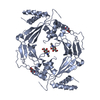 5j69C S: Starting model for refinement C: citing same article ( |
|---|---|
| Similar structure data |


-Links
 PDBj
PDBj- Assembly
| Deposited unit | 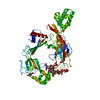
| ||||||||
|---|---|---|---|---|---|---|---|---|---|
| 1 |
| ||||||||
| Unit cell |
|
-Components
| #1: Protein | Mass: 65054.977 Da / Num. of mol.: 1 Source method: isolated from a genetically manipulated source Source: (gene. exp.) Homo sapiens (human) / Gene: ASTN2, KIAA0634 / Cell line (production host): HEK293 / Production host: Homo sapiens (human) / References: UniProt: O75129 |
|---|---|
| #2: Polysaccharide | alpha-D-mannopyranose-(1-2)-alpha-D-mannopyranose-(1-3)-[alpha-D-mannopyranose-(1-2)-alpha-D- ...alpha-D-mannopyranose-(1-2)-alpha-D-mannopyranose-(1-3)-[alpha-D-mannopyranose-(1-2)-alpha-D-mannopyranose-(1-6)]alpha-D-mannopyranose-(1-6)-[alpha-D-mannopyranose-(1-2)-alpha-D-mannopyranose-(1-3)]beta-D-mannopyranose-(1-4)-2-acetamido-2-deoxy-beta-D-glucopyranose-(1-4)-2-acetamido-2-deoxy-beta-D-glucopyranose Source method: isolated from a genetically manipulated source |
| #3: Sugar | ChemComp-NAG /   Type: D-saccharide, beta linking / Mass: 221.208 Da / Num. of mol.: 1 Type: D-saccharide, beta linking / Mass: 221.208 Da / Num. of mol.: 1Source method: isolated from a genetically manipulated source Formula: C8H15NO6 / Source: (gene. exp.) Homo sapiens (human) / Cell line (production host): HEK293 / Production host: Homo sapiens (human) |
| #4: Chemical | ChemComp-I3P / 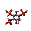  Mass: 420.096 Da / Num. of mol.: 1 Mass: 420.096 Da / Num. of mol.: 1Source method: isolated from a genetically manipulated source Formula: C6H15O15P3 / Source: (gene. exp.) Homo sapiens (human) / Cell line (production host): HEK293 / Production host: Homo sapiens (human) |
| Has protein modification | Y |
-Experimental details
-Experiment
| Experiment | Method: X-RAY DIFFRACTION / Number of used crystals: 1 |
|---|
- Sample preparation
| Crystal | Density Matthews: 6.31 Å3/Da / Density % sol: 80.51 % |
|---|---|
| Crystal grow | Temperature: 293 K / Method: vapor diffusion, sitting drop / Details: 0.1 M MES, pH 5.0 to 6.0, 5% PEG 6000 / PH range: 5-6 |
-Data collection
| Diffraction | Mean temperature: 100 K |
|---|---|
| Diffraction source | Source: SYNCHROTRON / Site: Diamond / Beamline: I03 / Wavelength: 0.97625 Å |
| Detector | Type: DECTRIS PILATUS 6M / Detector: PIXEL / Date: Oct 10, 2014 |
| Radiation | Protocol: SINGLE WAVELENGTH / Monochromatic (M) / Laue (L): M / Scattering type: x-ray |
| Radiation wavelength | Wavelength: 0.97625 Å / Relative weight: 1 |
| Reflection | Resolution: 5.22→85.802 Å / Num. obs: 6915 / % possible obs: 100 % / Redundancy: 23.8 % / Rmerge(I) obs: 0.12 / Net I/σ(I): 17.3 |
| Reflection shell | Highest resolution: 5.22 Å / Rmerge(I) obs: 3.047 |
- Processing
| Software |
| ||||||||||||||||||||||||||||
|---|---|---|---|---|---|---|---|---|---|---|---|---|---|---|---|---|---|---|---|---|---|---|---|---|---|---|---|---|---|
| Refinement | Method to determine structure: MOLECULAR REPLACEMENT Starting model: 5J67 Resolution: 5.221→85.802 Å / SU ML: 1.12 / Cross valid method: FREE R-VALUE / σ(F): 1.35 / Phase error: 50.1
| ||||||||||||||||||||||||||||
| Solvent computation | Shrinkage radii: 0.9 Å / VDW probe radii: 1.11 Å | ||||||||||||||||||||||||||||
| Refinement step | Cycle: LAST / Resolution: 5.221→85.802 Å
| ||||||||||||||||||||||||||||
| Refine LS restraints |
| ||||||||||||||||||||||||||||
| LS refinement shell |
|
