 Movie
Movie Controller

[English] 日本語
 Yorodumi
- PDB-5j14: Crystal structure of endoglycoceramidase I from Rhodococ-cus equi... -
+ Open data

- Basic information
| Entry | Database: PDB / ID: 5j14 | |||||||||
|---|---|---|---|---|---|---|---|---|---|---|
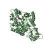
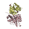
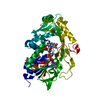
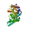
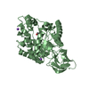
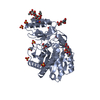
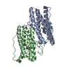
| Title | Crystal structure of endoglycoceramidase I from Rhodococ-cus equi in complex with GM3 | |||||||||
 Components Components | Putative secreted endoglycosylceramidase | |||||||||
 Keywords Keywords | HYDROLASE / Complex | |||||||||
| Function / homology |  Function and homology information Function and homology informationendoglycosylceramidase / endoglycosylceramidase activity / galactosylceramide catabolic process / polysaccharide catabolic process / extracellular region / membrane Similarity search - Function | |||||||||
| Biological species |  Rhodococcus equi 103S (bacteria) Rhodococcus equi 103S (bacteria) | |||||||||
| Method |  X-RAY DIFFRACTION / SYNCHROTRON / MOLECULAR REPLACEMENT / Resolution: 1.915 Å X-RAY DIFFRACTION / SYNCHROTRON / MOLECULAR REPLACEMENT / Resolution: 1.915 Å | |||||||||
 Authors Authors | Chen, L. | |||||||||
 Citation Citation | Journal: J. Biol. Chem. / Year: 2017 Title: Structural Insights into the Broad Substrate Specificity of a Novel Endoglycoceramidase I Belonging to a New Subfamily of GH5 Glycosidases Authors: Han, Y.B. / Chen, L.Q. / Li, Z. / Tan, Y.M. / Feng, Y. / Yang, G.Y. | |||||||||
| History |
|
- Structure visualization
| Structure viewer | Molecule: MolmilJmol/JSmol |
|---|
- Downloads & links
-Download
| PDBx/mmCIF format | 5j14.cif.gz | 201.9 KB | Display | PDBx/mmCIF format |
|---|---|---|---|---|
| PDB format | pdb5j14.ent.gz | 155.5 KB | Display | PDB format |
| PDBx/mmJSON format | 5j14.json.gz | Tree view | PDBx/mmJSON format | |
| Others |  Other downloads Other downloads |
-Validation report
| Summary document | 5j14_validation.pdf.gz | 1.7 MB | Display | wwPDB validaton report |
|---|---|---|---|---|
| Full document | 5j14_full_validation.pdf.gz | 1.7 MB | Display | |
| Data in XML | 5j14_validation.xml.gz | 40.7 KB | Display | |
| Data in CIF | 5j14_validation.cif.gz | 60.8 KB | Display | |
| Arichive directory | https://data.pdbj.org/pub/pdb/validation_reports/j1/5j14ftp://data.pdbj.org/pub/pdb/validation_reports/j1/5j14 | HTTPS FTP |
-Related structure data
| Related structure data | 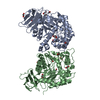 5ccuSC 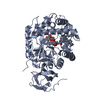 5j7zC C: citing same article ( S: Starting model for refinement |
|---|---|
| Similar structure data |


-Links
 PDBj
PDBj


- Assembly
| Deposited unit | 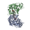
| ||||||||
|---|---|---|---|---|---|---|---|---|---|
| 1 | 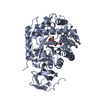
| ||||||||
| 2 | 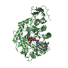
| ||||||||
| Unit cell |
|
-Components
-Protein , 1 types, 2 molecules AB
| #1: Protein | Mass: 53869.863 Da / Num. of mol.: 2 / Mutation: E347S Source method: isolated from a genetically manipulated source Source: (gene. exp.) Rhodococcus equi 103S (bacteria) / Strain: 103S / Gene: REQ_38260Production host: Strain (production host): BL21-Gold(DE3)pLysS AG / References: UniProt: E4W8N9, UniProt: A0A3S5YBC7*PLUS |
|---|
-Sugars , 2 types, 2 molecules
| #2: Polysaccharide | N-acetyl-alpha-neuraminic acid-(2-3)-beta-D-galactopyranose-(1-4)-beta-D-glucopyranose Source method: isolated from a genetically manipulated source |
|---|---|
| #3: Polysaccharide | beta-D-galactopyranose-(1-4)-beta-D-glucopyranose / beta-lactose  Source method: isolated from a genetically manipulated source Details: oligosaccharide / References: beta-lactose |
-Non-polymers , 3 types, 711 molecules 

| #4: Chemical |  Mass: 22.990 Da / Num. of mol.: 2 / Source method: obtained synthetically / Formula: Na Mass: 22.990 Da / Num. of mol.: 2 / Source method: obtained synthetically / Formula: Na#5: Chemical |  Mass: 565.954 Da / Num. of mol.: 2 / Source method: obtained synthetically / Formula: C36H71NO3 Mass: 565.954 Da / Num. of mol.: 2 / Source method: obtained synthetically / Formula: C36H71NO3#6: Water | ChemComp-HOH / | Mass: 18.015 Da / Num. of mol.: 707 / Source method: isolated from a natural source / Formula: H2O |
|---|
-Details
| Has protein modification | Y |
|---|
-Experimental details
-Experiment
| Experiment | Method: X-RAY DIFFRACTION / Number of used crystals: 1 |
|---|
- Sample preparation
| Crystal | Density Matthews: 2.4 Å3/Da / Density % sol: 48.78 % |
|---|---|
| Crystal grow | Temperature: 291 K / Method: vapor diffusion, hanging drop / Details: PEG 800, sodium hydroxide, ammonium chloride |
-Data collection
| Diffraction | Mean temperature: 100 K |
|---|---|
| Diffraction source | Source: SYNCHROTRON / Site: SSRF  / Beamline: BL17U / Wavelength: 0.979 Å / Beamline: BL17U / Wavelength: 0.979 Å |
| Detector | Type: ADSC QUANTUM 270 / Detector: CCD / Date: Oct 7, 2015 |
| Radiation | Protocol: SINGLE WAVELENGTH / Monochromatic (M) / Laue (L): M / Scattering type: x-ray |
| Radiation wavelength | Wavelength: 0.979 Å / Relative weight: 1 |
| Reflection | Resolution: 1.915→50 Å / Num. obs: 79432 / % possible obs: 97.4 % / Redundancy: 4.2 % / Rmerge(I) obs: 0.123 / Net I/σ(I): 11 |
- Processing
| Software |
| ||||||||||||||||
|---|---|---|---|---|---|---|---|---|---|---|---|---|---|---|---|---|---|
| Refinement | Method to determine structure: MOLECULAR REPLACEMENT Starting model: 5CCU Resolution: 1.915→50 Å / Cross valid method: THROUGHOUT
| ||||||||||||||||
| Refinement step | Cycle: LAST / Resolution: 1.915→50 Å
| ||||||||||||||||
| LS refinement shell | Resolution: 1.9153→1.9838 Å /
|
