 Movie
Movie Controller

[English] 日本語
 Yorodumi
- PDB-5ixt: The crystal structure of the Arabidopsis receptor kinase HAESA LR... -
+ Open data

- Basic information
| Entry | Database: PDB / ID: 5ixt | |||||||||
|---|---|---|---|---|---|---|---|---|---|---|
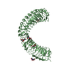
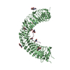
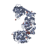
| Title | The crystal structure of the Arabidopsis receptor kinase HAESA LRR ectdomain in complex with a N-terminal extended IDA peptide hormone ligand. | |||||||||
 Components Components |
| |||||||||
 Keywords Keywords | SIGNALING PROTEIN / membrane receptor kinase / peptide hormone receptor / signaling complex / plant development / organ shedding | |||||||||
| Function / homology |  Function and homology information Function and homology informationlateral root morphogenesis / regulation of cell diameter / floral organ abscission / leaf abscission / response to ethylene / pectin catabolic process / apoplast / transmembrane receptor protein tyrosine kinase activity / receptor protein-tyrosine kinase / regulation of gene expression ...lateral root morphogenesis / regulation of cell diameter / floral organ abscission / leaf abscission / response to ethylene / pectin catabolic process / apoplast / transmembrane receptor protein tyrosine kinase activity / receptor protein-tyrosine kinase / regulation of gene expression / defense response to Gram-negative bacterium / protein kinase activity / non-specific serine/threonine protein kinase / signaling receptor binding / protein serine kinase activity / protein serine/threonine kinase activity / extracellular region / ATP binding / plasma membrane Similarity search - Function | |||||||||
| Biological species |  | |||||||||
| Method |  X-RAY DIFFRACTION / SYNCHROTRON / FOURIER SYNTHESIS / Resolution: 1.94 Å X-RAY DIFFRACTION / SYNCHROTRON / FOURIER SYNTHESIS / Resolution: 1.94 Å | |||||||||
 Authors Authors | Santiago, J. / Hothorn, M. | |||||||||
 Citation Citation | Journal: Elife / Year: 2016 Title: Mechanistic insight into a peptide hormone signaling complex mediating floral organ abscission. Authors: Santiago, J. / Brandt, B. / Wildhagen, M. / Hohmann, U. / Hothorn, L.A. / Butenko, M.A. / Hothorn, M. | |||||||||
| History |
|
- Structure visualization
| Structure viewer | Molecule: MolmilJmol/JSmol |
|---|
- Downloads & links
-Download
| PDBx/mmCIF format | 5ixt.cif.gz | 253.4 KB | Display | PDBx/mmCIF format |
|---|---|---|---|---|
| PDB format | pdb5ixt.ent.gz | 202.7 KB | Display | PDB format |
| PDBx/mmJSON format | 5ixt.json.gz | Tree view | PDBx/mmJSON format | |
| Others |  Other downloads Other downloads |
-Validation report
| Arichive directory | https://data.pdbj.org/pub/pdb/validation_reports/ix/5ixtftp://data.pdbj.org/pub/pdb/validation_reports/ix/5ixt | HTTPS FTP |
|---|
-Related structure data
| Related structure data |  5ixoSC  5ixqC  5iyvC 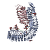 5iyxC S: Starting model for refinement C: citing same article ( |
|---|---|
| Similar structure data |


-Links
 PDBj
PDBj


- Assembly
| Deposited unit | 
| ||||||||
|---|---|---|---|---|---|---|---|---|---|
| 1 |
| ||||||||
| Unit cell |
|
-Components
-Protein / Protein/peptide , 2 types, 2 molecules AB
| #1: Protein | Mass: 67216.195 Da / Num. of mol.: 1 / Fragment: ectodomain, residues 20-620 Source method: isolated from a genetically manipulated source Source: (gene. exp.)  Trichoplusia ni (cabbage looper) / Strain (production host): BTI38 Trichoplusia ni (cabbage looper) / Strain (production host): BTI38References: UniProt: P47735, receptor protein-tyrosine kinase, non-specific serine/threonine protein kinase |
|---|---|
| #2: Protein/peptide | Mass: 1865.140 Da / Num. of mol.: 1 / Fragment: UNP residues 53-69 / Source method: obtained synthetically / Source: (synth.) |
-Sugars , 3 types, 7 molecules 
| #3: Polysaccharide | Source method: isolated from a genetically manipulated source #4: Polysaccharide | alpha-D-mannopyranose-(1-3)-beta-D-mannopyranose-(1-4)-2-acetamido-2-deoxy-beta-D-glucopyranose-(1- ...alpha-D-mannopyranose-(1-3)-beta-D-mannopyranose-(1-4)-2-acetamido-2-deoxy-beta-D-glucopyranose-(1-4)-2-acetamido-2-deoxy-beta-D-glucopyranose | Source method: isolated from a genetically manipulated source #5: Sugar |  Type: D-saccharide, beta linking / Mass: 221.208 Da / Num. of mol.: 3 Type: D-saccharide, beta linking / Mass: 221.208 Da / Num. of mol.: 3Source method: isolated from a genetically manipulated source Formula: C8H15NO6 |
|---|
-Non-polymers , 3 types, 43 molecules 


| #6: Chemical |  Mass: 24.305 Da / Num. of mol.: 2 / Source method: obtained synthetically / Formula: Mg Mass: 24.305 Da / Num. of mol.: 2 / Source method: obtained synthetically / Formula: Mg#7: Chemical | ChemComp-EDO / |  Mass: 62.068 Da / Num. of mol.: 1 / Source method: obtained synthetically / Formula: C2H6O2 Mass: 62.068 Da / Num. of mol.: 1 / Source method: obtained synthetically / Formula: C2H6O2#8: Water | ChemComp-HOH / | Mass: 18.015 Da / Num. of mol.: 40 / Source method: isolated from a natural source / Formula: H2O |
|---|
-Experimental details
-Experiment
| Experiment | Method: X-RAY DIFFRACTION / Number of used crystals: 1 |
|---|
- Sample preparation
| Crystal | Density Matthews: 2.69 Å3/Da / Density % sol: 54.25 % |
|---|---|
| Crystal grow | Temperature: 298 K / Method: vapor diffusion, hanging drop / pH: 4 / Details: 23% PEG 3350,0.2M MgCl2,0.1M citric acid pH4 |
-Data collection
| Diffraction | Mean temperature: 100 K |
|---|---|
| Diffraction source | Source: SYNCHROTRON / Site: SLS  / Beamline: X10SA / Wavelength: 0.99998 Å / Beamline: X10SA / Wavelength: 0.99998 Å |
| Detector | Type: DECTRIS PILATUS3 6M / Detector: PIXEL / Date: May 15, 2014 |
| Radiation | Protocol: SINGLE WAVELENGTH / Monochromatic (M) / Laue (L): M / Scattering type: x-ray |
| Radiation wavelength | Wavelength: 0.99998 Å / Relative weight: 1 |
| Reflection | Resolution: 1.94→128.97 Å / Num. obs: 54201 / % possible obs: 99.4 % / Observed criterion σ(F): -3 / Redundancy: 11.2 % / Biso Wilson estimate: 81.7 Å2 / CC1/2: 1 / Rsym value: 0.035 / Net I/σ(I): 20.9 |
| Reflection shell | Resolution: 1.94→2.06 Å / Redundancy: 11.1 % / Mean I/σ(I) obs: 2.4 / % possible all: 97.9 |
- Processing
| Software |
| ||||||||||||||||||||||||||||||||||||||||||||||||||||||||||||||||||||||||||||||||||||||||||||||||||||||||||||||||||||||||||||||||||||||||||||||||||||||||||||||||||||||||||||||||||||||
|---|---|---|---|---|---|---|---|---|---|---|---|---|---|---|---|---|---|---|---|---|---|---|---|---|---|---|---|---|---|---|---|---|---|---|---|---|---|---|---|---|---|---|---|---|---|---|---|---|---|---|---|---|---|---|---|---|---|---|---|---|---|---|---|---|---|---|---|---|---|---|---|---|---|---|---|---|---|---|---|---|---|---|---|---|---|---|---|---|---|---|---|---|---|---|---|---|---|---|---|---|---|---|---|---|---|---|---|---|---|---|---|---|---|---|---|---|---|---|---|---|---|---|---|---|---|---|---|---|---|---|---|---|---|---|---|---|---|---|---|---|---|---|---|---|---|---|---|---|---|---|---|---|---|---|---|---|---|---|---|---|---|---|---|---|---|---|---|---|---|---|---|---|---|---|---|---|---|---|---|---|---|---|---|
| Refinement | Method to determine structure: FOURIER SYNTHESIS Starting model: 5IXO Resolution: 1.94→128.97 Å / Cor.coef. Fo:Fc: 0.977 / Cor.coef. Fo:Fc free: 0.966 / SU B: 8.142 / SU ML: 0.106 / Cross valid method: THROUGHOUT / ESU R: 0.138 / ESU R Free: 0.126 / Details: HYDROGENS HAVE BEEN ADDED IN THE RIDING POSITIONS
| ||||||||||||||||||||||||||||||||||||||||||||||||||||||||||||||||||||||||||||||||||||||||||||||||||||||||||||||||||||||||||||||||||||||||||||||||||||||||||||||||||||||||||||||||||||||
| Solvent computation | Ion probe radii: 0.8 Å / Shrinkage radii: 0.8 Å / VDW probe radii: 1.2 Å | ||||||||||||||||||||||||||||||||||||||||||||||||||||||||||||||||||||||||||||||||||||||||||||||||||||||||||||||||||||||||||||||||||||||||||||||||||||||||||||||||||||||||||||||||||||||
| Displacement parameters | Biso mean: 80.66 Å2
| ||||||||||||||||||||||||||||||||||||||||||||||||||||||||||||||||||||||||||||||||||||||||||||||||||||||||||||||||||||||||||||||||||||||||||||||||||||||||||||||||||||||||||||||||||||||
| Refinement step | Cycle: 1 / Resolution: 1.94→128.97 Å
| ||||||||||||||||||||||||||||||||||||||||||||||||||||||||||||||||||||||||||||||||||||||||||||||||||||||||||||||||||||||||||||||||||||||||||||||||||||||||||||||||||||||||||||||||||||||
| Refine LS restraints |
|