 Movie
Movie Controller


+ Open data

- Basic information
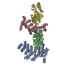
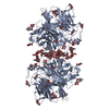
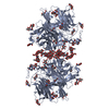
| Entry | Database: PDB / ID: 5imy | ||||||
|---|---|---|---|---|---|---|---|
| Title | Trapped Toxin | ||||||
 Components Components |
| ||||||
 Keywords Keywords | toxin/toxin receptor / toxin-toxin receptor complex | ||||||
| Function / homology |  Function and homology information Function and homology informationnegative regulation of activation of membrane attack complex / negative regulation of complement-dependent cytotoxicity / complement binding / negative regulation of complement activation / regulation of complement-dependent cytotoxicity / regulation of complement activation / Cargo concentration in the ER / COPII-mediated vesicle transport / cholesterol binding / tertiary granule membrane ...negative regulation of activation of membrane attack complex / negative regulation of complement-dependent cytotoxicity / complement binding / negative regulation of complement activation / regulation of complement-dependent cytotoxicity / regulation of complement activation / Cargo concentration in the ER / COPII-mediated vesicle transport / cholesterol binding / tertiary granule membrane / host cell membrane / specific granule membrane / transport vesicle / COPI-mediated anterograde transport / endoplasmic reticulum-Golgi intermediate compartment membrane / Regulation of Complement cascade / ER to Golgi transport vesicle membrane / protein sequestering activity / blood coagulation / toxin activity / killing of cells of another organism / vesicle / cell surface receptor signaling pathway / Golgi membrane / external side of plasma membrane / focal adhesion / Neutrophil degranulation / endoplasmic reticulum membrane / cell surface / : / extracellular exosome / extracellular region / membrane / plasma membrane Similarity search - Function | ||||||
| Biological species |  Gardnerella vaginalis (bacteria) Gardnerella vaginalis (bacteria) Homo sapiens (human) Homo sapiens (human) | ||||||
| Method |  X-RAY DIFFRACTION / SYNCHROTRON / MOLECULAR REPLACEMENT / Resolution: 2.4 Å X-RAY DIFFRACTION / SYNCHROTRON / MOLECULAR REPLACEMENT / Resolution: 2.4 Å | ||||||
 Authors Authors | Lawrence, S.L. / Morton, C.J. / Parker, M.W. | ||||||
 Citation Citation | Journal: Structure / Year: 2016 Title: Structural Basis for Receptor Recognition by the Human CD59-Responsive Cholesterol-Dependent Cytolysins. Authors: Lawrence, S.L. / Gorman, M.A. / Feil, S.C. / Mulhern, T.D. / Kuiper, M.J. / Ratner, A.J. / Tweten, R.K. / Morton, C.J. / Parker, M.W. | ||||||
| History |
|
- Structure visualization
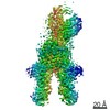
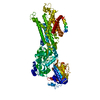
| Structure viewer | Molecule: MolmilJmol/JSmol |
|---|
- Downloads & links
-Download
| PDBx/mmCIF format | 5imy.cif.gz | 410.1 KB | Display | PDBx/mmCIF format |
|---|---|---|---|---|
| PDB format | pdb5imy.ent.gz | 339.9 KB | Display | PDB format |
| PDBx/mmJSON format | 5imy.json.gz | Tree view | PDBx/mmJSON format | |
| Others |  Other downloads Other downloads |
-Validation report
| Arichive directory | https://data.pdbj.org/pub/pdb/validation_reports/im/5imyftp://data.pdbj.org/pub/pdb/validation_reports/im/5imy | HTTPS FTP |
|---|
-Related structure data
| Related structure data |  5imtC  5imwC  1s3rS S: Starting model for refinement C: citing same article ( |
|---|---|
| Similar structure data |

-Links
 PDBj
PDBj


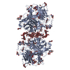
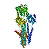
- Assembly
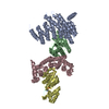
| Deposited unit | 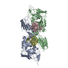
| ||||||||
|---|---|---|---|---|---|---|---|---|---|
| 1 |
| ||||||||
| Unit cell |
|
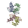
-Components
| #1: Protein | Mass: 54213.367 Da / Num. of mol.: 2 Source method: isolated from a genetically manipulated source Source: (gene. exp.) Gardnerella vaginalis (bacteria) / Gene: VLY, vly / Production host: #2: Protein | Mass: 9101.303 Da / Num. of mol.: 2 Source method: isolated from a genetically manipulated source Source: (gene. exp.) Homo sapiens (human) / Gene: CD59, MIC11, MIN1, MIN2, MIN3, MSK21 / Production host: #3: Water | ChemComp-HOH / |  Mass: 18.015 Da / Num. of mol.: 137 / Source method: isolated from a natural source / Formula: H2O Mass: 18.015 Da / Num. of mol.: 137 / Source method: isolated from a natural source / Formula: H2OHas protein modification | Y | |
|---|
-Experimental details
-Experiment
| Experiment | Method: X-RAY DIFFRACTION / Number of used crystals: 1 |
|---|
- Sample preparation
| Crystal | Density Matthews: 2.45 Å3/Da / Density % sol: 49.7 % |
|---|---|
| Crystal grow | Temperature: 293 K / Method: vapor diffusion, hanging drop Details: Ammonium acetate, PEG 10000, Bis-Tris, Magnesium chloride |
-Data collection
| Diffraction | Mean temperature: 100 K |
|---|---|
| Diffraction source | Source: SYNCHROTRON / Site: Australian Synchrotron  / Beamline: MX2 / Wavelength: 0.9537 Å / Beamline: MX2 / Wavelength: 0.9537 Å |
| Detector | Type: ADSC QUANTUM 315r / Detector: CCD / Date: Dec 4, 2015 |
| Radiation | Protocol: SINGLE WAVELENGTH / Monochromatic (M) / Laue (L): M / Scattering type: x-ray |
| Radiation wavelength | Wavelength: 0.9537 Å / Relative weight: 1 |
| Reflection | Resolution: 2.4→48.315 Å / Num. obs: 49244 / % possible obs: 99.9 % / Redundancy: 4.5 % / Net I/σ(I): 13.1 |
- Processing
| Software |
| |||||||||||||||||||||||||||||||||||||||||||||||||||||||||||||||||||||||||||||||||||||||||||||||||||||||||||||||||||||||||||||||||||||||||||||||||||||||||||||||||
|---|---|---|---|---|---|---|---|---|---|---|---|---|---|---|---|---|---|---|---|---|---|---|---|---|---|---|---|---|---|---|---|---|---|---|---|---|---|---|---|---|---|---|---|---|---|---|---|---|---|---|---|---|---|---|---|---|---|---|---|---|---|---|---|---|---|---|---|---|---|---|---|---|---|---|---|---|---|---|---|---|---|---|---|---|---|---|---|---|---|---|---|---|---|---|---|---|---|---|---|---|---|---|---|---|---|---|---|---|---|---|---|---|---|---|---|---|---|---|---|---|---|---|---|---|---|---|---|---|---|---|---|---|---|---|---|---|---|---|---|---|---|---|---|---|---|---|---|---|---|---|---|---|---|---|---|---|---|---|---|---|---|---|
| Refinement | Method to determine structure: MOLECULAR REPLACEMENT Starting model: 1S3R Resolution: 2.4→42.639 Å / SU ML: 0.4 / Cross valid method: FREE R-VALUE / σ(F): 1.34 / Phase error: 30.71
| |||||||||||||||||||||||||||||||||||||||||||||||||||||||||||||||||||||||||||||||||||||||||||||||||||||||||||||||||||||||||||||||||||||||||||||||||||||||||||||||||
| Solvent computation | Shrinkage radii: 0.9 Å / VDW probe radii: 1.11 Å | |||||||||||||||||||||||||||||||||||||||||||||||||||||||||||||||||||||||||||||||||||||||||||||||||||||||||||||||||||||||||||||||||||||||||||||||||||||||||||||||||
| Refinement step | Cycle: LAST / Resolution: 2.4→42.639 Å
| |||||||||||||||||||||||||||||||||||||||||||||||||||||||||||||||||||||||||||||||||||||||||||||||||||||||||||||||||||||||||||||||||||||||||||||||||||||||||||||||||
| Refine LS restraints |
| |||||||||||||||||||||||||||||||||||||||||||||||||||||||||||||||||||||||||||||||||||||||||||||||||||||||||||||||||||||||||||||||||||||||||||||||||||||||||||||||||
| LS refinement shell |
|
