 Movie
Movie Controller

[English] 日本語
 Yorodumi
- PDB-5imj: Crystal structure of a Z-ring associated protein from Escherichia coli -
+ Open data

- Basic information
| Entry | Database: PDB / ID: 5imj | ||||||
|---|---|---|---|---|---|---|---|
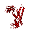
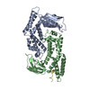
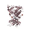
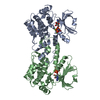
| Title | Crystal structure of a Z-ring associated protein from Escherichia coli | ||||||
 Components Components | Cell division protein ZapD | ||||||
 Keywords Keywords | CELL CYCLE / ZapD / cytokinesis / cell division / FtsZ | ||||||
| Function / homology |  Function and homology information Function and homology informationdivision septum assembly / FtsZ-dependent cytokinesis / cell division site / protein homodimerization activity / identical protein binding / cytosol Similarity search - Function | ||||||
| Biological species |  | ||||||
| Method |  X-RAY DIFFRACTION / SYNCHROTRON / MOLECULAR REPLACEMENT / Resolution: 3.1 Å X-RAY DIFFRACTION / SYNCHROTRON / MOLECULAR REPLACEMENT / Resolution: 3.1 Å | ||||||
 Authors Authors | Choi, H. / Yoon, H.J. / Lee, H.H. | ||||||
 Citation Citation | Journal: To Be Published Title: Crystal structure of ZapD, a positive regulator of Z-ring formation during bacterial cytokinesis Authors: Lee, H.H. / Choi, H. / Min, K.J. / Yoon, H.J. / Ha, J.M. | ||||||
| History |
|
- Structure visualization
| Structure viewer | Molecule: MolmilJmol/JSmol |
|---|
- Downloads & links
-Download
| PDBx/mmCIF format | 5imj.cif.gz | 112.1 KB | Display | PDBx/mmCIF format |
|---|---|---|---|---|
| PDB format | pdb5imj.ent.gz | 87.1 KB | Display | PDB format |
| PDBx/mmJSON format | 5imj.json.gz | Tree view | PDBx/mmJSON format | |
| Others |  Other downloads Other downloads |
-Validation report
| Arichive directory | https://data.pdbj.org/pub/pdb/validation_reports/im/5imjftp://data.pdbj.org/pub/pdb/validation_reports/im/5imj | HTTPS FTP |
|---|
-Related structure data
| Related structure data |  2oezS S: Starting model for refinement |
|---|---|
| Similar structure data |


-Links
 PDBj
PDBj- Assembly
| Deposited unit | 
| ||||||||
|---|---|---|---|---|---|---|---|---|---|
| 1 |
| ||||||||
| Unit cell |
| ||||||||
| Components on special symmetry positions |
|
-Components
| #1: Protein | Mass: 28192.396 Da / Num. of mol.: 2 / Fragment: UNP residues 2-247 Source method: isolated from a genetically manipulated source Source: (gene. exp.) Strain: K12 / Gene: zapD, yacF, b0102, JW0099 / Production host: #2: Chemical | ChemComp-SO4 /   Mass: 96.063 Da / Num. of mol.: 5 / Source method: obtained synthetically / Formula: SO4 Mass: 96.063 Da / Num. of mol.: 5 / Source method: obtained synthetically / Formula: SO4#3: Water | ChemComp-HOH / |  Mass: 18.015 Da / Num. of mol.: 41 / Source method: isolated from a natural source / Formula: H2O Mass: 18.015 Da / Num. of mol.: 41 / Source method: isolated from a natural source / Formula: H2O |
|---|
-Experimental details
-Experiment
| Experiment | Method: X-RAY DIFFRACTION / Number of used crystals: 1 |
|---|
- Sample preparation
| Crystal | Density Matthews: 3.27 Å3/Da / Density % sol: 62.42 % |
|---|---|
| Crystal grow | Temperature: 298 K / Method: vapor diffusion, sitting drop / Details: Lithium sulfate, HEPES pH 7.8, PEG400 |
-Data collection
| Diffraction | Mean temperature: 100 K |
|---|---|
| Diffraction source | Source: SYNCHROTRON / Site: PAL/PLS  / Beamline: 5C (4A) / Wavelength: 0.97952 Å / Beamline: 5C (4A) / Wavelength: 0.97952 Å |
| Detector | Type: ADSC QUANTUM 270 / Detector: CCD / Date: Jun 2, 2013 |
| Radiation | Protocol: SINGLE WAVELENGTH / Monochromatic (M) / Laue (L): M / Scattering type: x-ray |
| Radiation wavelength | Wavelength: 0.97952 Å / Relative weight: 1 |
| Reflection | Resolution: 2.95→50 Å / Num. obs: 15284 / % possible obs: 99.3 % / Redundancy: 9.1 % / Rmerge(I) obs: 0.505 / Rsym value: 0.505 / Net I/σ(I): 33.4 |
| Reflection shell | Resolution: 2.95→3 Å |
- Processing
| Software |
| ||||||||||||||||||||||||||||||||||||||||||||||||||||||||||||||||||||||||||||||||||||||||||||||||||||||||||||||||||||||||||||||||||||||||||||||||||||||||||||||||||||||||||||||||||||||
|---|---|---|---|---|---|---|---|---|---|---|---|---|---|---|---|---|---|---|---|---|---|---|---|---|---|---|---|---|---|---|---|---|---|---|---|---|---|---|---|---|---|---|---|---|---|---|---|---|---|---|---|---|---|---|---|---|---|---|---|---|---|---|---|---|---|---|---|---|---|---|---|---|---|---|---|---|---|---|---|---|---|---|---|---|---|---|---|---|---|---|---|---|---|---|---|---|---|---|---|---|---|---|---|---|---|---|---|---|---|---|---|---|---|---|---|---|---|---|---|---|---|---|---|---|---|---|---|---|---|---|---|---|---|---|---|---|---|---|---|---|---|---|---|---|---|---|---|---|---|---|---|---|---|---|---|---|---|---|---|---|---|---|---|---|---|---|---|---|---|---|---|---|---|---|---|---|---|---|---|---|---|---|---|
| Refinement | Method to determine structure: MOLECULAR REPLACEMENT Starting model: 2OEZ Resolution: 3.1→35.43 Å / Cor.coef. Fo:Fc: 0.941 / Cor.coef. Fo:Fc free: 0.902 / SU B: 21.347 / SU ML: 0.373 / Cross valid method: THROUGHOUT / ESU R Free: 0.109 / Details: HYDROGENS HAVE BEEN ADDED IN THE RIDING POSITIONS
| ||||||||||||||||||||||||||||||||||||||||||||||||||||||||||||||||||||||||||||||||||||||||||||||||||||||||||||||||||||||||||||||||||||||||||||||||||||||||||||||||||||||||||||||||||||||
| Solvent computation | Ion probe radii: 0.8 Å / Shrinkage radii: 0.8 Å / VDW probe radii: 1.2 Å | ||||||||||||||||||||||||||||||||||||||||||||||||||||||||||||||||||||||||||||||||||||||||||||||||||||||||||||||||||||||||||||||||||||||||||||||||||||||||||||||||||||||||||||||||||||||
| Displacement parameters | Biso mean: 94.516 Å2
| ||||||||||||||||||||||||||||||||||||||||||||||||||||||||||||||||||||||||||||||||||||||||||||||||||||||||||||||||||||||||||||||||||||||||||||||||||||||||||||||||||||||||||||||||||||||
| Refinement step | Cycle: 1 / Resolution: 3.1→35.43 Å
| ||||||||||||||||||||||||||||||||||||||||||||||||||||||||||||||||||||||||||||||||||||||||||||||||||||||||||||||||||||||||||||||||||||||||||||||||||||||||||||||||||||||||||||||||||||||
| Refine LS restraints |
|