 Movie
Movie Controller


+ Open data

- Basic information
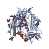
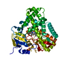
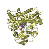
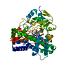
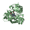
| Entry | Database: PDB / ID: 5iki | ||||||
|---|---|---|---|---|---|---|---|
| Title | CYP106A2 WITH SUBSTRATE ABIETIC ACID | ||||||
 Components Components | Cytochrome P450(MEG) | ||||||
 Keywords Keywords | OXIDOREDUCTASE / MONO-OXYGENASE / CYTOCHROME P450 / 15-BETA-HYDROXYLASE | ||||||
| Function / homology |  Function and homology information Function and homology informationsteroid 15beta-monooxygenase / Oxidoreductases; Acting on paired donors, with incorporation or reduction of molecular oxygen; Miscellaneous / oxidoreductase activity, acting on paired donors, with incorporation or reduction of molecular oxygen / monooxygenase activity / iron ion binding / heme binding / cytoplasm Similarity search - Function | ||||||
| Biological species |  Bacillus megaterium (bacteria) Bacillus megaterium (bacteria) | ||||||
| Method |  X-RAY DIFFRACTION / SYNCHROTRON / MOLECULAR REPLACEMENT / Resolution: 2.4 Å X-RAY DIFFRACTION / SYNCHROTRON / MOLECULAR REPLACEMENT / Resolution: 2.4 Å | ||||||
 Authors Authors | Janocha, S. / Carius, Y. / Bernhardt, R. / Lancaster, C.R.D. | ||||||
 Citation Citation | Journal: Chembiochem / Year: 2016 Title: Crystal Structure of CYP106A2 in Substrate-Free and Substrate-Bound Form. Authors: Janocha, S. / Carius, Y. / Hutter, M. / Lancaster, C.R. / Bernhardt, R. #1: Journal: Chembiochem / Year: 2011 Title: Identification of CYP106A2 as a regioselective allylic bacterial diterpene hydroxylase. Authors: Bleif, S. / Hannemann, F. / Lisurek, M. / Von Kries, J.P. / Zapp, J. / Dietzen, M. / Antes, I. / Bernhardt, R. | ||||||
| History |
|
- Structure visualization
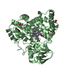
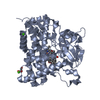
| Structure viewer | Molecule: MolmilJmol/JSmol |
|---|
- Downloads & links
-Download
| PDBx/mmCIF format | 5iki.cif.gz | 171.7 KB | Display | PDBx/mmCIF format |
|---|---|---|---|---|
| PDB format | pdb5iki.ent.gz | 135.2 KB | Display | PDB format |
| PDBx/mmJSON format | 5iki.json.gz | Tree view | PDBx/mmJSON format | |
| Others |  Other downloads Other downloads |
-Validation report
| Arichive directory | https://data.pdbj.org/pub/pdb/validation_reports/ik/5ikiftp://data.pdbj.org/pub/pdb/validation_reports/ik/5iki | HTTPS FTP |
|---|
-Related structure data
| Related structure data |  4yt3C  1f4tS C: citing same article ( S: Starting model for refinement |
|---|---|
| Similar structure data |

-Links
 PDBj
PDBj
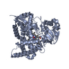
- Assembly
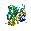
| Deposited unit | 
| ||||||||||||||||||
|---|---|---|---|---|---|---|---|---|---|---|---|---|---|---|---|---|---|---|---|
| 1 | 
| ||||||||||||||||||
| 2 | 
| ||||||||||||||||||
| Unit cell |
| ||||||||||||||||||
| Noncrystallographic symmetry (NCS) | NCS domain:
NCS domain segments: Component-ID: _ / Ens-ID: 1 / Beg auth comp-ID: VAL / Beg label comp-ID: VAL / End auth comp-ID: ARG / End label comp-ID: ARG / Refine code: _ / Auth seq-ID: 4 - 409 / Label seq-ID: 4 - 409
|
-Components
| #1: Protein | Mass: 47015.477 Da / Num. of mol.: 2 Source method: isolated from a genetically manipulated source Source: (gene. exp.) Bacillus megaterium (bacteria) / Gene: cyp106A2 / Plasmid: PKKHC / Production host: References: UniProt: Q06069, Oxidoreductases; Acting on paired donors, with incorporation or reduction of molecular oxygen; Miscellaneous, steroid 15beta-monooxygenase #2: Chemical |   Mass: 616.487 Da / Num. of mol.: 2 / Source method: obtained synthetically / Formula: C34H32FeN4O4 Mass: 616.487 Da / Num. of mol.: 2 / Source method: obtained synthetically / Formula: C34H32FeN4O4#3: Chemical | ChemComp-A9H / |   Mass: 302.451 Da / Num. of mol.: 1 / Source method: obtained synthetically / Formula: C20H30O2 Mass: 302.451 Da / Num. of mol.: 1 / Source method: obtained synthetically / Formula: C20H30O2#4: Water | ChemComp-HOH / |  Mass: 18.015 Da / Num. of mol.: 54 / Source method: isolated from a natural source / Formula: H2O Mass: 18.015 Da / Num. of mol.: 54 / Source method: isolated from a natural source / Formula: H2O |
|---|
-Experimental details
-Experiment
| Experiment | Method: X-RAY DIFFRACTION / Number of used crystals: 1 |
|---|
- Sample preparation
| Crystal | Density Matthews: 2.28 Å3/Da / Density % sol: 45.99 % |
|---|---|
| Crystal grow | Temperature: 291 K / Method: vapor diffusion, hanging drop / pH: 4.2 Details: PEG 4000 AMMONIUM ACETATE SODIUM ACETATE TRIHYDRATE |
-Data collection
| Diffraction | Mean temperature: 100 K |
|---|---|
| Diffraction source | Source: SYNCHROTRON / Site: ESRF  / Beamline: ID14-4 / Wavelength: 0.9393 Å / Beamline: ID14-4 / Wavelength: 0.9393 Å |
| Detector | Type: ADSC QUANTUM 315r / Detector: CCD / Date: May 17, 2012 |
| Radiation | Protocol: SINGLE WAVELENGTH / Monochromatic (M) / Laue (L): M / Scattering type: x-ray |
| Radiation wavelength | Wavelength: 0.9393 Å / Relative weight: 1 |
| Reflection | Resolution: 2.4→78.98 Å / Num. obs: 32407 / % possible obs: 94.93 % / Redundancy: 3.82 % / Biso Wilson estimate: 46.2 Å2 / Rmerge(I) obs: 0.07 / Net I/σ(I): 6.39 |
| Reflection shell | Resolution: 2.4→2.53 Å / Redundancy: 4.11 % / Rmerge(I) obs: 0.22 / Mean I/σ(I) obs: 2.94 / % possible all: 95.3 |
- Processing
| Software |
| ||||||||||||||||||||||||||||||||||||||||||||||||||||||||||||||||||||||||||||||||||||||||||||||||||||||||||||||||||||||||||||||||||||||||||||||||||||||||||||||||||||||||||||||||||||||
|---|---|---|---|---|---|---|---|---|---|---|---|---|---|---|---|---|---|---|---|---|---|---|---|---|---|---|---|---|---|---|---|---|---|---|---|---|---|---|---|---|---|---|---|---|---|---|---|---|---|---|---|---|---|---|---|---|---|---|---|---|---|---|---|---|---|---|---|---|---|---|---|---|---|---|---|---|---|---|---|---|---|---|---|---|---|---|---|---|---|---|---|---|---|---|---|---|---|---|---|---|---|---|---|---|---|---|---|---|---|---|---|---|---|---|---|---|---|---|---|---|---|---|---|---|---|---|---|---|---|---|---|---|---|---|---|---|---|---|---|---|---|---|---|---|---|---|---|---|---|---|---|---|---|---|---|---|---|---|---|---|---|---|---|---|---|---|---|---|---|---|---|---|---|---|---|---|---|---|---|---|---|---|---|
| Refinement | Method to determine structure: MOLECULAR REPLACEMENT Starting model: 1F4T Resolution: 2.4→78.73 Å / Cor.coef. Fo:Fc: 0.96 / Cor.coef. Fo:Fc free: 0.932 / SU B: 14.552 / SU ML: 0.309 / Cross valid method: THROUGHOUT / ESU R: 0.67 / ESU R Free: 0.304 / Details: HYDROGENS HAVE BEEN ADDED IN THE RIDING POSITIONS
| ||||||||||||||||||||||||||||||||||||||||||||||||||||||||||||||||||||||||||||||||||||||||||||||||||||||||||||||||||||||||||||||||||||||||||||||||||||||||||||||||||||||||||||||||||||||
| Solvent computation | Ion probe radii: 0.8 Å / Shrinkage radii: 0.8 Å / VDW probe radii: 1.2 Å | ||||||||||||||||||||||||||||||||||||||||||||||||||||||||||||||||||||||||||||||||||||||||||||||||||||||||||||||||||||||||||||||||||||||||||||||||||||||||||||||||||||||||||||||||||||||
| Displacement parameters | Biso mean: 63.362 Å2
| ||||||||||||||||||||||||||||||||||||||||||||||||||||||||||||||||||||||||||||||||||||||||||||||||||||||||||||||||||||||||||||||||||||||||||||||||||||||||||||||||||||||||||||||||||||||
| Refinement step | Cycle: 1 / Resolution: 2.4→78.73 Å
| ||||||||||||||||||||||||||||||||||||||||||||||||||||||||||||||||||||||||||||||||||||||||||||||||||||||||||||||||||||||||||||||||||||||||||||||||||||||||||||||||||||||||||||||||||||||
| Refine LS restraints |
|