Mass: 18.015 Da / Num. of mol.: 133 / Source method: isolated from a natural source / Formula: H2O
Has protein modification
Y
Sequence details
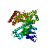
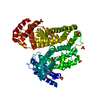
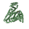
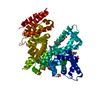
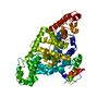
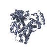
The authors state that in contrast with the previously deposited in PDB structures of Equus ...The authors state that in contrast with the previously deposited in PDB structures of Equus caballus SA (PDB IDs: 3V08, 4J2V, 4OT2, 4F5U, and 4F5T), a single point mutation, R561A, is observed. The long arginine side chain cannot be modeled in this position due to steric clashes with the nearby disulfide bond connecting Cys567 and Cys558 and a symmetry-related copy of the molecule. Moreover, there is no 2mFo-DFc omit map supporting placement of the side chain. Protein was purified from natural source, therefore there may be naturally occurring mutation. According to the NCBI database, this mutation is characteristic for Equus ferus przewalskii, a rare subspecies of wild horse from central Asia (accession code: XP_008524663.1). However it is possible that there is an error in the Equus caballus SA sequence, or the observed mutation naturally occurs in that species.
-
Experimental details
-
Experiment
Experiment
Method: X-RAY DIFFRACTION / Number of used crystals: 1
-
Sample preparation
Crystal
Density Matthews: 2.69 Å3/Da / Density % sol: 54.28 %
Crystal grow
Temperature: 289 K / Method: vapor diffusion, hanging drop / pH: 7.4 Details: 1 ul of 30 mg/ml protein in 10 mM Tris pH 7.5 and 150 mM NaCl buffer was mixed with 1 ul of the well condition (0.2 M Li2SO4, 0.1 M Tris:HCl pH 7.4, 2.0 M (NH4)2SO4, 5 mM ZnCl2) and ...Details: 1 ul of 30 mg/ml protein in 10 mM Tris pH 7.5 and 150 mM NaCl buffer was mixed with 1 ul of the well condition (0.2 M Li2SO4, 0.1 M Tris:HCl pH 7.4, 2.0 M (NH4)2SO4, 5 mM ZnCl2) and equilibrated against well solution in 15 Well Crystallization Plate (Qiagen)
Resolution: 2.4→50.01 Å / Cor.coef. Fo:Fc: 0.967 / Cor.coef. Fo:Fc free: 0.937 / SU B: 20.102 / SU ML: 0.218 / SU R Cruickshank DPI: 0.3451 / Cross valid method: THROUGHOUT / σ(F): 0 / ESU R: 0.345 / ESU R Free: 0.245 Details: U VALUES : WITH TLS ADDED HYDROGENS HAVE BEEN ADDED IN THE RIDING POSITIONS
Rfactor
Num. reflection
% reflection
Selection details
Rfree
0.2354
1348
5 %
RANDOM
Rwork
0.1748
-
-
-
obs
0.1777
25677
99.65 %
-
Solvent computation
Ion probe radii: 0.8 Å / Shrinkage radii: 0.8 Å / VDW probe radii: 1.2 Å
In the structure databanks used in Yorodumi, some data are registered as the other names, "COVID-19 virus" and "2019-nCoV". Here are the details of the virus and the list of structure data.
Jan 31, 2019. EMDB accession codes are about to change! (news from PDBe EMDB page)
EMDB accession codes are about to change! (news from PDBe EMDB page)
The allocation of 4 digits for EMDB accession codes will soon come to an end. Whilst these codes will remain in use, new EMDB accession codes will include an additional digit and will expand incrementally as the available range of codes is exhausted. The current 4-digit format prefixed with “EMD-” (i.e. EMD-XXXX) will advance to a 5-digit format (i.e. EMD-XXXXX), and so on. It is currently estimated that the 4-digit codes will be depleted around Spring 2019, at which point the 5-digit format will come into force.
The EM Navigator/Yorodumi systems omit the EMD- prefix.
Related info.:Q: What is EMD? / ID/Accession-code notation in Yorodumi/EM Navigator
Yorodumi is a browser for structure data from EMDB, PDB, SASBDB, etc.
This page is also the successor to EM Navigator detail page, and also detail information page/front-end page for Omokage search.
The word "yorodu" (or yorozu) is an old Japanese word meaning "ten thousand". "mi" (miru) is to see.
Related info.:EMDB / PDB / SASBDB / Comparison of 3 databanks / Yorodumi Search / Aug 31, 2016. New EM Navigator & Yorodumi / Yorodumi Papers / Jmol/JSmol / Function and homology information / Changes in new EM Navigator and Yorodumi
 Movie
Movie Controller

 Yorodumi
 Open data
 Basic information
 Components
 Keywords
 Function and homology information
 X-RAY DIFFRACTION /
  Authors
 United States, 3items
  Citation
 Structure visualization
 Downloads & links
 Other downloads


 PDBj
PDBj


 Assembly

 Mass: 65.409 Da / Num. of mol.: 1 / Source method: obtained synthetically / Formula: Zn
 Mass: 96.063 Da / Num. of mol.: 1 / Source method: obtained synthetically / Formula: SO4
 Mass: 18.015 Da / Num. of mol.: 133 / Source method: isolated from a natural source / Formula: H2O
 Sample preparation
 Processing