 Movie
Movie Controller

[English] 日本語
 Yorodumi
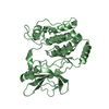
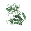
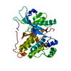
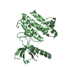
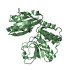
- PDB-5idv: Structure of the nucleotide binding domain of an ABC transporter ... -
+ Open data

- Basic information
| Entry | Database: PDB / ID: 5idv | ||||||
|---|---|---|---|---|---|---|---|
| Title | Structure of the nucleotide binding domain of an ABC transporter MsbA from Acinetobacter baumannii | ||||||
 Components Components | Lipid A export ATP-binding/permease protein MsbA | ||||||
 Keywords Keywords | TRANSPORT PROTEIN / SSGCID / Acinetobacter baumannii / MsbA / nucleotide binding domain / ATP-binding / Lipid A export / Structural Genomics / Seattle Structural Genomics Center for Infectious Disease | ||||||
| Function / homology | P-loop containing nucleotide triphosphate hydrolases / Rossmann fold / 3-Layer(aba) Sandwich / Alpha Beta / :  Function and homology information Function and homology information | ||||||
| Biological species |  Acinetobacter baumannii AB5075 (bacteria) Acinetobacter baumannii AB5075 (bacteria) | ||||||
| Method |  X-RAY DIFFRACTION / SYNCHROTRON / MOLECULAR REPLACEMENT / molecular replacement / Resolution: 1.45 Å X-RAY DIFFRACTION / SYNCHROTRON / MOLECULAR REPLACEMENT / molecular replacement / Resolution: 1.45 Å | ||||||
 Authors Authors | Seattle Structural Genomics Center for Infectious Disease (SSGCID) | ||||||
 Citation Citation | Journal: to be published Title: Structure of the nucleotide binding domain of an ABC transporter MsbA from Acinetobacter baumannii Authors: Mayclin, S.J. / Lorimer, D.D. / Edwards, T.E. | ||||||
| History |
|
- Structure visualization
| Structure viewer | Molecule: MolmilJmol/JSmol |
|---|
- Downloads & links
-Download
| PDBx/mmCIF format | 5idv.cif.gz | 127.5 KB | Display | PDBx/mmCIF format |
|---|---|---|---|---|
| PDB format | pdb5idv.ent.gz | 97.8 KB | Display | PDB format |
| PDBx/mmJSON format | 5idv.json.gz | Tree view | PDBx/mmJSON format | |
| Others |  Other downloads Other downloads |
-Validation report
| Arichive directory | https://data.pdbj.org/pub/pdb/validation_reports/id/5idvftp://data.pdbj.org/pub/pdb/validation_reports/id/5idv | HTTPS FTP |
|---|
-Related structure data
| Related structure data | 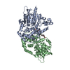 4qlaS S: Starting model for refinement |
|---|---|
| Similar structure data | |
| Other databases |


-Links
 PDBj
PDBj- Assembly
| Deposited unit | 
| ||||||||
|---|---|---|---|---|---|---|---|---|---|
| 1 |
| ||||||||
| 2 | 
| ||||||||
| Unit cell |
| ||||||||
| Components on special symmetry positions |
|
-Components
| #1: Protein | Mass: 31028.783 Da / Num. of mol.: 1 / Fragment: AcbaC.18886.a.B2 (UNP residues 305-575) Source method: isolated from a genetically manipulated source Source: (gene. exp.) Acinetobacter baumannii AB5075 (bacteria)Gene: msbA, APC40_11520 / Plasmid: AcbaC.18886.a.B2 / Production host: References: UniProt: A0A0Q2LVW2, Hydrolases; Acting on acid anhydrides; Acting on acid anhydrides to catalyse transmembrane movement of substances | ||
|---|---|---|---|
| #2: Chemical |   Mass: 62.068 Da / Num. of mol.: 2 / Source method: obtained synthetically / Formula: C2H6O2 Mass: 62.068 Da / Num. of mol.: 2 / Source method: obtained synthetically / Formula: C2H6O2#3: Water | ChemComp-HOH / |  Mass: 18.015 Da / Num. of mol.: 287 / Source method: isolated from a natural source / Formula: H2O Mass: 18.015 Da / Num. of mol.: 287 / Source method: isolated from a natural source / Formula: H2O |
-Experimental details
-Experiment
| Experiment | Method: X-RAY DIFFRACTION / Number of used crystals: 1 |
|---|
- Sample preparation
| Crystal | Density Matthews: 2.44 Å3/Da / Density % sol: 49.59 % / Mosaicity: 0.22 ° |
|---|---|
| Crystal grow | Temperature: 290 K / Method: vapor diffusion, sitting drop / pH: 7.5 Details: JCSG+ A8 (267191a8): 200mM Ammonium formate, 20% PEG3350; protein conc. 17.3mg/mL; 20% ethylene glycol cryo; puck aak7-1 |
-Data collection
| Diffraction | Mean temperature: 100 K | ||||||||||||||||||||||||||||||||||||||||||||||||||||||||||||||||||||||||||||||||||||||||||||||||||||||||||||||||||||||||||||||
|---|---|---|---|---|---|---|---|---|---|---|---|---|---|---|---|---|---|---|---|---|---|---|---|---|---|---|---|---|---|---|---|---|---|---|---|---|---|---|---|---|---|---|---|---|---|---|---|---|---|---|---|---|---|---|---|---|---|---|---|---|---|---|---|---|---|---|---|---|---|---|---|---|---|---|---|---|---|---|---|---|---|---|---|---|---|---|---|---|---|---|---|---|---|---|---|---|---|---|---|---|---|---|---|---|---|---|---|---|---|---|---|---|---|---|---|---|---|---|---|---|---|---|---|---|---|---|---|
| Diffraction source | Source: SYNCHROTRON / Site: APS  / Beamline: 21-ID-F / Wavelength: 0.97872 Å / Beamline: 21-ID-F / Wavelength: 0.97872 Å | ||||||||||||||||||||||||||||||||||||||||||||||||||||||||||||||||||||||||||||||||||||||||||||||||||||||||||||||||||||||||||||||
| Detector | Type: RAYONIX MX-225 / Detector: CCD / Date: Nov 20, 2015 | ||||||||||||||||||||||||||||||||||||||||||||||||||||||||||||||||||||||||||||||||||||||||||||||||||||||||||||||||||||||||||||||
| Radiation | Monochromator: Diamond [111] / Protocol: SINGLE WAVELENGTH / Monochromatic (M) / Laue (L): M / Scattering type: x-ray | ||||||||||||||||||||||||||||||||||||||||||||||||||||||||||||||||||||||||||||||||||||||||||||||||||||||||||||||||||||||||||||||
| Radiation wavelength | Wavelength: 0.97872 Å / Relative weight: 1 | ||||||||||||||||||||||||||||||||||||||||||||||||||||||||||||||||||||||||||||||||||||||||||||||||||||||||||||||||||||||||||||||
| Reflection | Resolution: 1.45→52.613 Å / Num. obs: 53798 / % possible obs: 99.7 % / Observed criterion σ(I): -3 / Redundancy: 6.14 % / Biso Wilson estimate: 23.521 Å2 / CC1/2: 1 / Rmerge(I) obs: 0.04 / Net I/σ(I): 25 | ||||||||||||||||||||||||||||||||||||||||||||||||||||||||||||||||||||||||||||||||||||||||||||||||||||||||||||||||||||||||||||||
| Reflection shell |
|
-Phasing
| Phasing | Method: molecular replacement |
|---|
- Processing
| Software |
| ||||||||||||||||||||||||
|---|---|---|---|---|---|---|---|---|---|---|---|---|---|---|---|---|---|---|---|---|---|---|---|---|---|
| Refinement | Method to determine structure: MOLECULAR REPLACEMENT Starting model: 4QLA Resolution: 1.45→50 Å / SU ML: 0.12 / Cross valid method: FREE R-VALUE / σ(F): 1.35 / Phase error: 16.7
| ||||||||||||||||||||||||
| Solvent computation | Shrinkage radii: 0.9 Å / VDW probe radii: 1.11 Å | ||||||||||||||||||||||||
| Displacement parameters | Biso max: 63.98 Å2 / Biso mean: 22.9851 Å2 / Biso min: 9.95 Å2 | ||||||||||||||||||||||||
| Refinement step | Cycle: final / Resolution: 1.45→50 Å
|
