 Movie
Movie Controller

[English] 日本語
 Yorodumi
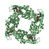
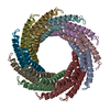
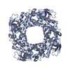
- PDB-5i72: Crystal structure of the oligomeric form of the Lassa virus matri... -
+ Open data

- Basic information
| Entry | Database: PDB / ID: 5i72 | ||||||
|---|---|---|---|---|---|---|---|
| Title | Crystal structure of the oligomeric form of the Lassa virus matrix protein Z | ||||||
 Components Components | RING finger protein Z | ||||||
 Keywords Keywords | VIRAL PROTEIN / arenavirus / Lassa virus / matrix / Z / oligomer | ||||||
| Function / homology |  Function and homology information Function and homology informationviral budding via host ESCRT complex / viral budding from plasma membrane / virion component / host cell perinuclear region of cytoplasm / host cell plasma membrane / RNA binding / zinc ion binding / membrane Similarity search - Function | ||||||
| Biological species |  Lassa virus Lassa virus | ||||||
| Method |  X-RAY DIFFRACTION / SYNCHROTRON / SAD / Resolution: 2.9 Å X-RAY DIFFRACTION / SYNCHROTRON / SAD / Resolution: 2.9 Å | ||||||
 Authors Authors | Hastie, K. / Zandonatti, M. / Liu, T. / Li, S. / Woods Jr, V. / Saphire, E.O. | ||||||
| Funding support |  United States, 1items United States, 1items
| ||||||
 Citation Citation | Journal: J.Virol. / Year: 2016 Title: Crystal Structure of the Oligomeric Form of Lassa Virus Matrix Protein Z. Authors: Hastie, K.M. / Zandonatti, M. / Liu, T. / Li, S. / Woods, V.L. / Saphire, E.O. | ||||||
| History |
|
- Structure visualization
| Structure viewer | Molecule: MolmilJmol/JSmol |
|---|
- Downloads & links
-Download
| PDBx/mmCIF format | 5i72.cif.gz | 35.1 KB | Display | PDBx/mmCIF format |
|---|---|---|---|---|
| PDB format | pdb5i72.ent.gz | 23 KB | Display | PDB format |
| PDBx/mmJSON format | 5i72.json.gz | Tree view | PDBx/mmJSON format | |
| Others |  Other downloads Other downloads |
-Validation report
| Summary document | 5i72_validation.pdf.gz | 433.9 KB | Display | wwPDB validaton report |
|---|---|---|---|---|
| Full document | 5i72_full_validation.pdf.gz | 434.4 KB | Display | |
| Data in XML | 5i72_validation.xml.gz | 6 KB | Display | |
| Data in CIF | 5i72_validation.cif.gz | 7.1 KB | Display | |
| Arichive directory | https://data.pdbj.org/pub/pdb/validation_reports/i7/5i72ftp://data.pdbj.org/pub/pdb/validation_reports/i7/5i72 | HTTPS FTP |
-Related structure data
| Similar structure data |
|---|


-Links
 PDBj
PDBj- Assembly
| Deposited unit | 
| |||||||||||||||||||||||||||
|---|---|---|---|---|---|---|---|---|---|---|---|---|---|---|---|---|---|---|---|---|---|---|---|---|---|---|---|---|
| 1 | x 6
| |||||||||||||||||||||||||||
| Unit cell |
| |||||||||||||||||||||||||||
| Noncrystallographic symmetry (NCS) | NCS domain:
NCS domain segments:
|
-Components
| #1: Protein | Mass: 6085.327 Da / Num. of mol.: 2 / Fragment: unp residues 25-77 Source method: isolated from a genetically manipulated source Source: (gene. exp.) Lassa virus (strain Mouse/Sierra Leone/Josiah/1976)Strain: Mouse/Sierra Leone/Josiah/1976 / Production host:  #2: Chemical | ChemComp-ZN /   Mass: 65.409 Da / Num. of mol.: 4 / Source method: obtained synthetically / Formula: Zn Mass: 65.409 Da / Num. of mol.: 4 / Source method: obtained synthetically / Formula: Zn |
|---|
-Experimental details
-Experiment
| Experiment | Method: X-RAY DIFFRACTION / Number of used crystals: 1 |
|---|
- Sample preparation
| Crystal | Density Matthews: 4.49 Å3/Da / Density % sol: 72.61 % |
|---|---|
| Crystal grow | Temperature: 293 K / Method: vapor diffusion, hanging drop Details: 300-400mM ammonium sulfate, 100mM HEPES pH 7.5 and 17% PEG 3350 |
-Data collection
| Diffraction | Mean temperature: 100 K | ||||||||||||||||||||||||||||||||||||||||||||||||||||||||||||||||||||||||||||||||||||||||||||||||||||||||||||||
|---|---|---|---|---|---|---|---|---|---|---|---|---|---|---|---|---|---|---|---|---|---|---|---|---|---|---|---|---|---|---|---|---|---|---|---|---|---|---|---|---|---|---|---|---|---|---|---|---|---|---|---|---|---|---|---|---|---|---|---|---|---|---|---|---|---|---|---|---|---|---|---|---|---|---|---|---|---|---|---|---|---|---|---|---|---|---|---|---|---|---|---|---|---|---|---|---|---|---|---|---|---|---|---|---|---|---|---|---|---|---|---|
| Diffraction source | Source: SYNCHROTRON / Site: ALS / Beamline: 5.0.2 / Wavelength: 1.2827 Å | ||||||||||||||||||||||||||||||||||||||||||||||||||||||||||||||||||||||||||||||||||||||||||||||||||||||||||||||
| Detector | Type: 3 x 3 CCD array (ADSC Q315R) / Detector: CCD / Date: Feb 10, 2010 | ||||||||||||||||||||||||||||||||||||||||||||||||||||||||||||||||||||||||||||||||||||||||||||||||||||||||||||||
| Radiation | Protocol: SINGLE WAVELENGTH / Monochromatic (M) / Laue (L): M / Scattering type: x-ray | ||||||||||||||||||||||||||||||||||||||||||||||||||||||||||||||||||||||||||||||||||||||||||||||||||||||||||||||
| Radiation wavelength | Wavelength: 1.2827 Å / Relative weight: 1 | ||||||||||||||||||||||||||||||||||||||||||||||||||||||||||||||||||||||||||||||||||||||||||||||||||||||||||||||
| Reflection | Resolution: 2.9→38.221 Å / Num. obs: 9325 / % possible obs: 99.8 % / Redundancy: 4.8 % / Biso Wilson estimate: 86.73 Å2 / Rmerge(I) obs: 0.074 / Rrim(I) all: 0.083 / Χ2: 0.93 / Net I/σ(I): 10.2 / Num. measured all: 47129 / Scaling rejects: 2357 | ||||||||||||||||||||||||||||||||||||||||||||||||||||||||||||||||||||||||||||||||||||||||||||||||||||||||||||||
| Reflection shell | Diffraction-ID: 1
|
- Processing
| Software |
| ||||||||||||||||||||||||||||
|---|---|---|---|---|---|---|---|---|---|---|---|---|---|---|---|---|---|---|---|---|---|---|---|---|---|---|---|---|---|
| Refinement | Method to determine structure: SAD / Resolution: 2.9→38.221 Å / SU ML: 0.12 / Cross valid method: FREE R-VALUE / σ(F): 0.94 / Phase error: 25.82 / Stereochemistry target values: ML
| ||||||||||||||||||||||||||||
| Solvent computation | Shrinkage radii: 0.9 Å / VDW probe radii: 1.11 Å / Solvent model: FLAT BULK SOLVENT MODEL | ||||||||||||||||||||||||||||
| Displacement parameters | Biso max: 162.25 Å2 / Biso mean: 89.7017 Å2 / Biso min: 52.25 Å2 | ||||||||||||||||||||||||||||
| Refinement step | Cycle: final / Resolution: 2.9→38.221 Å
| ||||||||||||||||||||||||||||
| Refine LS restraints |
| ||||||||||||||||||||||||||||
| Refine LS restraints NCS |
| ||||||||||||||||||||||||||||
| LS refinement shell | Refine-ID: X-RAY DIFFRACTION / Total num. of bins used: 3
|
