 Movie
Movie Controller


+ Open data

- Basic information
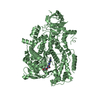
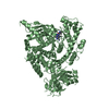
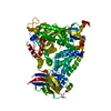
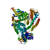
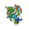
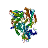
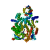
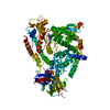
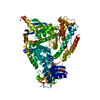
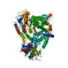
| Entry | Database: PDB / ID: 5i6u | ||||||
|---|---|---|---|---|---|---|---|
| Title | The crystal structure of PI3Kdelta with compound 32 | ||||||
 Components Components | Phosphatidylinositol 4,5-bisphosphate 3-kinase catalytic subunit delta isoform | ||||||
 Keywords Keywords | TRANSFERASE/TRANSFERASE INHIBITOR / kinase / p110 / kinase inhibitor / TRANSFERASE-TRANSFERASE INHIBITOR complex | ||||||
| Function / homology |  Function and homology information Function and homology informationCo-stimulation by ICOS / Erythropoietin activates Phosphoinositide-3-kinase (PI3K) / CD28 dependent PI3K/Akt signaling / Interleukin receptor SHC signaling / Synthesis of PIPs at the plasma membrane / PIP3 activates AKT signaling / Antigen activates B Cell Receptor (BCR) leading to generation of second messengers / Regulation of signaling by CBL / PI5P, PP2A and IER3 Regulate PI3K/AKT Signaling / Interleukin-3, Interleukin-5 and GM-CSF signaling ...Co-stimulation by ICOS / Erythropoietin activates Phosphoinositide-3-kinase (PI3K) / CD28 dependent PI3K/Akt signaling / Interleukin receptor SHC signaling / Synthesis of PIPs at the plasma membrane / PIP3 activates AKT signaling / Antigen activates B Cell Receptor (BCR) leading to generation of second messengers / Regulation of signaling by CBL / PI5P, PP2A and IER3 Regulate PI3K/AKT Signaling / Interleukin-3, Interleukin-5 and GM-CSF signaling / RET signaling / positive regulation of epithelial tube formation / positive regulation of neutrophil apoptotic process / phosphatidylinositol 3-kinase complex / 1-phosphatidylinositol-4-phosphate 3-kinase activity / phosphatidylinositol 3-kinase complex, class IA / phosphatidylinositol-4,5-bisphosphate 3-kinase / 1-phosphatidylinositol-4,5-bisphosphate 3-kinase activity / phosphatidylinositol 3-kinase / phosphatidylinositol-3-phosphate biosynthetic process / 1-phosphatidylinositol-3-kinase activity / vascular endothelial growth factor signaling pathway / phosphatidylinositol-mediated signaling / B cell homeostasis / B cell activation / homeostasis of number of cells / defense response to fungus / positive regulation of endothelial cell proliferation / positive regulation of endothelial cell migration / phosphatidylinositol 3-kinase/protein kinase B signal transduction / chemotaxis / positive regulation of angiogenesis / cell migration / adaptive immune response / cell differentiation / cell surface receptor signaling pathway / positive regulation of cell migration / inflammatory response / negative regulation of gene expression / innate immune response / positive regulation of gene expression / ATP binding / plasma membrane / cytoplasm / cytosol Similarity search - Function | ||||||
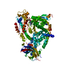
| Biological species |  | ||||||
| Method |  X-RAY DIFFRACTION / SYNCHROTRON / MOLECULAR REPLACEMENT / Resolution: 2.842 Å X-RAY DIFFRACTION / SYNCHROTRON / MOLECULAR REPLACEMENT / Resolution: 2.842 Å | ||||||
 Authors Authors | Somoza, J.R. / Villasenor, A.G. | ||||||
 Citation Citation | Journal: To Be Published Title: The crystal structure of PI3Kdelta with compound 32 Authors: Somoza, J.R. / Villasenor, A.G. #1: Journal: Acta Crystallogr. D Biol. Crystallogr. / Year: 2012 Title: Towards automated crystallographic structure refinement with phenix.refine. Authors: Afonine, P.V. / Grosse-Kunstleve, R.W. / Echols, N. / Headd, J.J. / Moriarty, N.W. / Mustyakimov, M. / Terwilliger, T.C. / Urzhumtsev, A. / Zwart, P.H. / Adams, P.D. #2: Journal: Acta Crystallogr D Biol Crystallogr / Year: 2010 Title: PHENIX: a comprehensive Python-based system for macromolecular structure solution. Authors: Paul D Adams / Pavel V Afonine / Gábor Bunkóczi / Vincent B Chen / Ian W Davis / Nathaniel Echols / Jeffrey J Headd / Li-Wei Hung / Gary J Kapral / Ralf W Grosse-Kunstleve / Airlie J McCoy ...Authors: Paul D Adams / Pavel V Afonine / Gábor Bunkóczi / Vincent B Chen / Ian W Davis / Nathaniel Echols / Jeffrey J Headd / Li-Wei Hung / Gary J Kapral / Ralf W Grosse-Kunstleve / Airlie J McCoy / Nigel W Moriarty / Robert Oeffner / Randy J Read / David C Richardson / Jane S Richardson / Thomas C Terwilliger / Peter H Zwart /  Abstract: Macromolecular X-ray crystallography is routinely applied to understand biological processes at a molecular level. However, significant time and effort are still required to solve and complete many ...Macromolecular X-ray crystallography is routinely applied to understand biological processes at a molecular level. However, significant time and effort are still required to solve and complete many of these structures because of the need for manual interpretation of complex numerical data using many software packages and the repeated use of interactive three-dimensional graphics. PHENIX has been developed to provide a comprehensive system for macromolecular crystallographic structure solution with an emphasis on the automation of all procedures. This has relied on the development of algorithms that minimize or eliminate subjective input, the development of algorithms that automate procedures that are traditionally performed by hand and, finally, the development of a framework that allows a tight integration between the algorithms. | ||||||
| History |
|
- Structure visualization
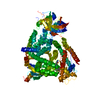
| Structure viewer | Molecule: MolmilJmol/JSmol |
|---|
- Downloads & links
-Download
| PDBx/mmCIF format | 5i6u.cif.gz | 222.5 KB | Display | PDBx/mmCIF format |
|---|---|---|---|---|
| PDB format | pdb5i6u.ent.gz | 139 KB | Display | PDB format |
| PDBx/mmJSON format | 5i6u.json.gz | Tree view | PDBx/mmJSON format | |
| Others |  Other downloads Other downloads |
-Validation report
| Arichive directory | https://data.pdbj.org/pub/pdb/validation_reports/i6/5i6uftp://data.pdbj.org/pub/pdb/validation_reports/i6/5i6u | HTTPS FTP |
|---|
-Related structure data
| Related structure data |  4xe0S S: Starting model for refinement |
|---|---|
| Similar structure data |


-Links
 PDBj
PDBj


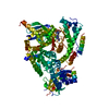
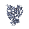
- Assembly
| Deposited unit | 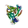
| ||||||||||||
|---|---|---|---|---|---|---|---|---|---|---|---|---|---|
| 1 |
| ||||||||||||
| Unit cell |
|
-Components
| #1: Protein | Mass: 107766.609 Da / Num. of mol.: 1 / Fragment: UNP residues 106-1043 Source method: isolated from a genetically manipulated source Source: (gene. exp.)   Spodoptera frugiperda (fall armyworm) Spodoptera frugiperda (fall armyworm)References: UniProt: O35904, phosphatidylinositol-4,5-bisphosphate 3-kinase |
|---|---|
| #2: Chemical | ChemComp-68R /   Mass: 482.924 Da / Num. of mol.: 1 / Source method: obtained synthetically / Formula: C25H19ClN8O Mass: 482.924 Da / Num. of mol.: 1 / Source method: obtained synthetically / Formula: C25H19ClN8O |
-Experimental details
-Experiment
| Experiment | Method: X-RAY DIFFRACTION / Number of used crystals: 1 |
|---|
- Sample preparation
| Crystal | Density Matthews: 2.45 Å3/Da / Density % sol: 49.84 % |
|---|---|
| Crystal grow | Temperature: 293 K / Method: vapor diffusion Details: Crystallization protocol can be found in Somoza et al. (2015). JBC 290, pp. 8439-8446. |
-Data collection
| Diffraction | Mean temperature: 100 K | |||||||||||||||||||||||||||||||||||||||||||||||||||||||||||||||||||||||||||||||||||||||||||||||||||||||||
|---|---|---|---|---|---|---|---|---|---|---|---|---|---|---|---|---|---|---|---|---|---|---|---|---|---|---|---|---|---|---|---|---|---|---|---|---|---|---|---|---|---|---|---|---|---|---|---|---|---|---|---|---|---|---|---|---|---|---|---|---|---|---|---|---|---|---|---|---|---|---|---|---|---|---|---|---|---|---|---|---|---|---|---|---|---|---|---|---|---|---|---|---|---|---|---|---|---|---|---|---|---|---|---|---|---|---|
| Diffraction source | Source: SYNCHROTRON / Site: ALS / Beamline: 5.0.1 / Wavelength: 0.977 Å | |||||||||||||||||||||||||||||||||||||||||||||||||||||||||||||||||||||||||||||||||||||||||||||||||||||||||
| Detector | Type: ADSC QUANTUM 315r / Detector: CCD / Date: Jun 7, 2012 | |||||||||||||||||||||||||||||||||||||||||||||||||||||||||||||||||||||||||||||||||||||||||||||||||||||||||
| Radiation | Protocol: SINGLE WAVELENGTH / Monochromatic (M) / Laue (L): M / Scattering type: x-ray | |||||||||||||||||||||||||||||||||||||||||||||||||||||||||||||||||||||||||||||||||||||||||||||||||||||||||
| Radiation wavelength | Wavelength: 0.977 Å / Relative weight: 1 | |||||||||||||||||||||||||||||||||||||||||||||||||||||||||||||||||||||||||||||||||||||||||||||||||||||||||
| Reflection | Resolution: 2.84→30 Å / Num. obs: 24148 / % possible obs: 97.4 % / Redundancy: 3.4 % / Biso Wilson estimate: 57.003 Å2 / Rmerge(I) obs: 0.132 / Net I/av σ(I): 11.891 / Net I/σ(I): 7.3 | |||||||||||||||||||||||||||||||||||||||||||||||||||||||||||||||||||||||||||||||||||||||||||||||||||||||||
| Reflection shell |
|
- Processing
| Software |
| |||||||||||||||||||||||||||||||||||||||||||||||||||||||||||||||||||||||||||||||||||||||||||||||||||||||||
|---|---|---|---|---|---|---|---|---|---|---|---|---|---|---|---|---|---|---|---|---|---|---|---|---|---|---|---|---|---|---|---|---|---|---|---|---|---|---|---|---|---|---|---|---|---|---|---|---|---|---|---|---|---|---|---|---|---|---|---|---|---|---|---|---|---|---|---|---|---|---|---|---|---|---|---|---|---|---|---|---|---|---|---|---|---|---|---|---|---|---|---|---|---|---|---|---|---|---|---|---|---|---|---|---|---|---|
| Refinement | Method to determine structure: MOLECULAR REPLACEMENT Starting model: 4XE0 Resolution: 2.842→29.662 Å / SU ML: 0.489828277376 / Cross valid method: FREE R-VALUE / σ(F): 1.341 / Phase error: 33.764564163
| |||||||||||||||||||||||||||||||||||||||||||||||||||||||||||||||||||||||||||||||||||||||||||||||||||||||||
| Solvent computation | Shrinkage radii: 0.9 Å / VDW probe radii: 1.11 Å | |||||||||||||||||||||||||||||||||||||||||||||||||||||||||||||||||||||||||||||||||||||||||||||||||||||||||
| Displacement parameters | Biso mean: 53.7675292773 Å2 | |||||||||||||||||||||||||||||||||||||||||||||||||||||||||||||||||||||||||||||||||||||||||||||||||||||||||
| Refinement step | Cycle: LAST / Resolution: 2.842→29.662 Å
| |||||||||||||||||||||||||||||||||||||||||||||||||||||||||||||||||||||||||||||||||||||||||||||||||||||||||
| Refine LS restraints |
| |||||||||||||||||||||||||||||||||||||||||||||||||||||||||||||||||||||||||||||||||||||||||||||||||||||||||
| LS refinement shell |
|
