 Movie
Movie Controller


+ Open data

- Basic information
| Entry | Database: PDB / ID: 5i3s | ||||||
|---|---|---|---|---|---|---|---|
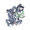
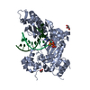
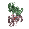
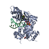
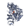
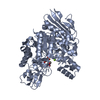
| Title | Crystal structure of Staphylococcal IMPase-II | ||||||
 Components Components | Inositol monophosphatase family protein | ||||||
 Keywords Keywords | HYDROLASE / IMPase / FIG superfamily / sugar phosphatase fold / staphylococcus aureus | ||||||
| Function / homology |  Function and homology information Function and homology informationinositol monophosphate 1-phosphatase activity / inositol metabolic process / signal transduction / metal ion binding Similarity search - Function | ||||||
| Biological species |   Staphylococcus aureus (bacteria) Staphylococcus aureus (bacteria) | ||||||
| Method |  X-RAY DIFFRACTION / MOLECULAR REPLACEMENT / molecular replacement / Resolution: 2.2 Å X-RAY DIFFRACTION / MOLECULAR REPLACEMENT / molecular replacement / Resolution: 2.2 Å | ||||||
 Authors Authors | Dutta, A. / Bhattacharyya, S. / Dutta, D. / Das, A.K. | ||||||
 Citation Citation | Journal: To Be Published Title: Crystal structure of Staphylococcal IMPase-II Authors: Dutta, A. / Bhattacharyya, S. / Dutta, D. / Das, A.K. | ||||||
| History |
|
- Structure visualization
| Structure viewer | Molecule: MolmilJmol/JSmol |
|---|
- Downloads & links
-Download
| PDBx/mmCIF format | 5i3s.cif.gz | 210.9 KB | Display | PDBx/mmCIF format |
|---|---|---|---|---|
| PDB format | pdb5i3s.ent.gz | 167.5 KB | Display | PDB format |
| PDBx/mmJSON format | 5i3s.json.gz | Tree view | PDBx/mmJSON format | |
| Others |  Other downloads Other downloads |
-Validation report
| Arichive directory | https://data.pdbj.org/pub/pdb/validation_reports/i3/5i3sftp://data.pdbj.org/pub/pdb/validation_reports/i3/5i3s | HTTPS FTP |
|---|
-Related structure data
| Related structure data |  3qflS S: Starting model for refinement |
|---|---|
| Similar structure data |


-Links
 PDBj
PDBj- Assembly
| Deposited unit | 
| ||||||||
|---|---|---|---|---|---|---|---|---|---|
| 1 | 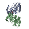
| ||||||||
| 2 | 
| ||||||||
| Unit cell |
|
-Components
| #1: Protein | Mass: 30487.246 Da / Num. of mol.: 4 Source method: isolated from a genetically manipulated source Source: (gene. exp.) Staphylococcus aureus (strain MSSA476) (bacteria)Strain: MSSA476 / Gene: SAS1042 / Plasmid: pQE30 / Production host: #2: Chemical | ChemComp-PO4 /   Mass: 94.971 Da / Num. of mol.: 4 / Source method: obtained synthetically / Formula: PO4 Mass: 94.971 Da / Num. of mol.: 4 / Source method: obtained synthetically / Formula: PO4#3: Water | ChemComp-HOH / |  Mass: 18.015 Da / Num. of mol.: 404 / Source method: isolated from a natural source / Formula: H2O Mass: 18.015 Da / Num. of mol.: 404 / Source method: isolated from a natural source / Formula: H2O |
|---|
-Experimental details
-Experiment
| Experiment | Method: X-RAY DIFFRACTION / Number of used crystals: 1 |
|---|
- Sample preparation
| Crystal | Density Matthews: 2.36 Å3/Da / Density % sol: 47.96 % |
|---|---|
| Crystal grow | Temperature: 298 K / Method: vapor diffusion, hanging drop / pH: 6 Details: 0.3M Ammonium dihydrogen phosphate, 0.1M Bis-Tris pH 6.0, 25% (w/v) PEG 3350 |
-Data collection
| Diffraction | Mean temperature: 100 K | |||||||||||||||||||||||||||||||||||||||||||||||||||||||||||||||||||||||||||||||||||||||||||||||||||||||||||||||||||||||||
|---|---|---|---|---|---|---|---|---|---|---|---|---|---|---|---|---|---|---|---|---|---|---|---|---|---|---|---|---|---|---|---|---|---|---|---|---|---|---|---|---|---|---|---|---|---|---|---|---|---|---|---|---|---|---|---|---|---|---|---|---|---|---|---|---|---|---|---|---|---|---|---|---|---|---|---|---|---|---|---|---|---|---|---|---|---|---|---|---|---|---|---|---|---|---|---|---|---|---|---|---|---|---|---|---|---|---|---|---|---|---|---|---|---|---|---|---|---|---|---|---|---|---|
| Diffraction source | Source: ROTATING ANODE / Type: RIGAKU MICROMAX-007 HF / Wavelength: 1.5418 Å | |||||||||||||||||||||||||||||||||||||||||||||||||||||||||||||||||||||||||||||||||||||||||||||||||||||||||||||||||||||||||
| Detector | Type: RIGAKU RAXIS IV++ / Detector: IMAGE PLATE / Date: Aug 13, 2013 | |||||||||||||||||||||||||||||||||||||||||||||||||||||||||||||||||||||||||||||||||||||||||||||||||||||||||||||||||||||||||
| Radiation | Monochromator: varimax / Protocol: SINGLE WAVELENGTH / Monochromatic (M) / Laue (L): M / Scattering type: x-ray | |||||||||||||||||||||||||||||||||||||||||||||||||||||||||||||||||||||||||||||||||||||||||||||||||||||||||||||||||||||||||
| Radiation wavelength | Wavelength: 1.5418 Å / Relative weight: 1 | |||||||||||||||||||||||||||||||||||||||||||||||||||||||||||||||||||||||||||||||||||||||||||||||||||||||||||||||||||||||||
| Reflection | Resolution: 2.202→78.349 Å / Num. all: 56328 / Num. obs: 56328 / % possible obs: 98.4 % / Redundancy: 7.6 % / Rpim(I) all: 0.046 / Rrim(I) all: 0.127 / Rsym value: 0.118 / Net I/av σ(I): 6.298 / Net I/σ(I): 16.7 / Num. measured all: 426223 | |||||||||||||||||||||||||||||||||||||||||||||||||||||||||||||||||||||||||||||||||||||||||||||||||||||||||||||||||||||||||
| Reflection shell | Diffraction-ID: 1 / Rejects: _
|
-Phasing
| Phasing | Method: molecular replacement | ||||||
|---|---|---|---|---|---|---|---|
| Phasing MR |
|
- Processing
| Software |
| |||||||||||||||||||||||||||||||||||||||||||||||||||||||||||||||||||||||||||
|---|---|---|---|---|---|---|---|---|---|---|---|---|---|---|---|---|---|---|---|---|---|---|---|---|---|---|---|---|---|---|---|---|---|---|---|---|---|---|---|---|---|---|---|---|---|---|---|---|---|---|---|---|---|---|---|---|---|---|---|---|---|---|---|---|---|---|---|---|---|---|---|---|---|---|---|---|
| Refinement | Method to determine structure: MOLECULAR REPLACEMENT Starting model: 3QFL Resolution: 2.2→78.35 Å / Cor.coef. Fo:Fc: 0.901 / Cor.coef. Fo:Fc free: 0.863 / WRfactor Rfree: 0.2411 / WRfactor Rwork: 0.1929 / FOM work R set: 0.7645 / SU B: 7.586 / SU ML: 0.184 / SU R Cruickshank DPI: 0.3309 / SU Rfree: 0.2519 / Cross valid method: THROUGHOUT / σ(F): 0 / ESU R: 0.331 / ESU R Free: 0.252 / Stereochemistry target values: MAXIMUM LIKELIHOOD Details: HYDROGENS HAVE BEEN ADDED IN THE RIDING POSITIONS U VALUES : REFINED INDIVIDUALLY
| |||||||||||||||||||||||||||||||||||||||||||||||||||||||||||||||||||||||||||
| Solvent computation | Ion probe radii: 0.8 Å / Shrinkage radii: 0.8 Å / VDW probe radii: 1.2 Å / Solvent model: MASK | |||||||||||||||||||||||||||||||||||||||||||||||||||||||||||||||||||||||||||
| Displacement parameters | Biso max: 84.14 Å2 / Biso mean: 27.908 Å2 / Biso min: 7.72 Å2
| |||||||||||||||||||||||||||||||||||||||||||||||||||||||||||||||||||||||||||
| Refinement step | Cycle: final / Resolution: 2.2→78.35 Å
| |||||||||||||||||||||||||||||||||||||||||||||||||||||||||||||||||||||||||||
| Refine LS restraints |
| |||||||||||||||||||||||||||||||||||||||||||||||||||||||||||||||||||||||||||
| LS refinement shell | Resolution: 2.202→2.259 Å / Total num. of bins used: 20
|
