 Movie
Movie Controller


+ Open data

- Basic information
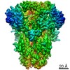
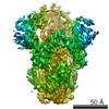
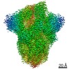
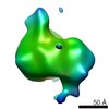
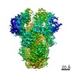
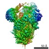
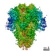
| Entry | Database: PDB / ID: 5i08 | ||||||
|---|---|---|---|---|---|---|---|
| Title | Prefusion structure of a human coronavirus spike protein | ||||||
 Components Components | Spike glycoprotein,Foldon chimera | ||||||
 Keywords Keywords | VIRAL PROTEIN / coronavirus / glycoprotein / prefusion | ||||||
| Function / homology |  Function and homology information Function and homology informationvirion component / host cell endoplasmic reticulum-Golgi intermediate compartment membrane / receptor-mediated virion attachment to host cell / endocytosis involved in viral entry into host cell / fusion of virus membrane with host plasma membrane / fusion of virus membrane with host endosome membrane / viral envelope / host cell plasma membrane / virion membrane / membrane / identical protein binding Similarity search - Function | ||||||
| Biological species |  Human coronavirus HKU1Enterobacteria phage T4 (virus) Human coronavirus HKU1Enterobacteria phage T4 (virus) | ||||||
| Method | ELECTRON MICROSCOPY / single particle reconstruction / cryo EM / Resolution: 4.04 Å | ||||||
 Authors Authors | Kirchdoerfer, R.N. / Cottrell, C.A. / Wang, N. / Pallesen, J. / Yassine, H.M. / Turner, H.L. / Corbett, K.S. / Graham, B.S. / McLellan, J.S. / Ward, A.B. | ||||||
 Citation Citation | Journal: Nature / Year: 2016 Title: Pre-fusion structure of a human coronavirus spike protein. Authors: Robert N Kirchdoerfer / Christopher A Cottrell / Nianshuang Wang / Jesper Pallesen / Hadi M Yassine / Hannah L Turner / Kizzmekia S Corbett / Barney S Graham / Jason S McLellan / Andrew B Ward /  Abstract: HKU1 is a human betacoronavirus that causes mild yet prevalent respiratory disease, and is related to the zoonotic SARS and MERS betacoronaviruses, which have high fatality rates and pandemic ...HKU1 is a human betacoronavirus that causes mild yet prevalent respiratory disease, and is related to the zoonotic SARS and MERS betacoronaviruses, which have high fatality rates and pandemic potential. Cell tropism and host range is determined in part by the coronavirus spike (S) protein, which binds cellular receptors and mediates membrane fusion. As the largest known class I fusion protein, its size and extensive glycosylation have hindered structural studies of the full ectodomain, thus preventing a molecular understanding of its function and limiting development of effective interventions. Here we present the 4.0 Å resolution structure of the trimeric HKU1 S protein determined using single-particle cryo-electron microscopy. In the pre-fusion conformation, the receptor-binding subunits, S1, rest above the fusion-mediating subunits, S2, preventing their conformational rearrangement. Surprisingly, the S1 C-terminal domains are interdigitated and form extensive quaternary interactions that occlude surfaces known in other coronaviruses to bind protein receptors. These features, along with the location of the two protease sites known to be important for coronavirus entry, provide a structural basis to support a model of membrane fusion mediated by progressive S protein destabilization through receptor binding and proteolytic cleavage. These studies should also serve as a foundation for the structure-based design of betacoronavirus vaccine immunogens. | ||||||
| History |
|
- Structure visualization
| Movie |
Movie viewer |
|---|---|
| Structure viewer | Molecule: MolmilJmol/JSmol |
- Downloads & links
-Download
| PDBx/mmCIF format | 5i08.cif.gz | 527 KB | Display | PDBx/mmCIF format |
|---|---|---|---|---|
| PDB format | pdb5i08.ent.gz | 404.8 KB | Display | PDB format |
| PDBx/mmJSON format | 5i08.json.gz | Tree view | PDBx/mmJSON format | |
| Others |  Other downloads Other downloads |
-Validation report
| Arichive directory | https://data.pdbj.org/pub/pdb/validation_reports/i0/5i08ftp://data.pdbj.org/pub/pdb/validation_reports/i0/5i08 | HTTPS FTP |
|---|
-Related structure data
| Related structure data | 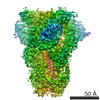 8069MC 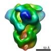 8066C 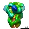 8067C 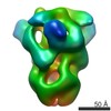 8068C M: map data used to model this data C: citing same article ( |
|---|---|
| Similar structure data |
-Links
 PDBj
PDBj

- Assembly
| Deposited unit | 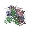
|
|---|---|
| 1 |
|
-Components
| #1: Protein | Mass: 144295.891 Da / Num. of mol.: 3 Mutation: R751G, R752G, K753S, R754G, R755S,R751G, R752G, K753S, R754G, R755S Source method: isolated from a genetically manipulated source Source: (gene. exp.) Human coronavirus HKU1 (isolate N5), (gene. exp.) Enterobacteria phage T4 (virus)Strain: isolate N5 / Gene: S, 3, wac / Plasmid: pVRC-8400 / Cell line (production host): FreeStyle 293F / Production host:  Homo sapiens (human) / References: UniProt: Q0ZME7, UniProt: P10104 Homo sapiens (human) / References: UniProt: Q0ZME7, UniProt: P10104Has protein modification | Y | |
|---|
-Experimental details
-Experiment
| Experiment | Method: ELECTRON MICROSCOPY |
|---|---|
| EM experiment | Aggregation state: PARTICLE / 3D reconstruction method: single particle reconstruction |
- Sample preparation
| Component | Name: HKU1 spike with attached foldon domain and mutated furin-cleavage site Type: COMPLEX / Entity ID: all / Source: RECOMBINANT | |||||||||||||||
|---|---|---|---|---|---|---|---|---|---|---|---|---|---|---|---|---|
| Molecular weight | Value: 0.42 MDa / Experimental value: NO | |||||||||||||||
| Source (natural) | Organism: Human coronavirus HKU1 (isolate N5) | |||||||||||||||
| Source (recombinant) | Organism: Homo sapiens (human) / Cell: FreeStyle 293F / Plasmid: pVRC8400 | |||||||||||||||
| Buffer solution | pH: 8 | |||||||||||||||
| Buffer component |
| |||||||||||||||
| Specimen | Conc.: 0.27 mg/ml / Embedding applied: NO / Shadowing applied: NO / Staining applied: NO / Vitrification applied: YES | |||||||||||||||
| EM staining | Type: NEGATIVE Details: 3 uL sample was applied to grid for 30 seconds and then blotted. Grids were stained with 3 uL 1% uranyl formate for 60 seconds followed by blotting. Material: uranyl formate | |||||||||||||||
| Specimen support | Grid material: COPPER / Grid mesh size: 400 divisions/in. / Grid type: EMS CF-2/2-4C C-Flat | |||||||||||||||
| Vitrification | Instrument: HOMEMADE PLUNGER / Cryogen name: ETHANE Details: 3 uL sample was applied to grid, blotted, and plunged into liquid ethane. |
- Electron microscopy imaging
| Experimental equipment |  Model: Titan Krios / Image courtesy: FEI Company |
|---|---|
| Microscopy | Model: FEI TITAN KRIOS |
| Electron gun | Electron source:  FIELD EMISSION GUN / Accelerating voltage: 300 kV / Illumination mode: FLOOD BEAM FIELD EMISSION GUN / Accelerating voltage: 300 kV / Illumination mode: FLOOD BEAM |
| Electron lens | Mode: BRIGHT FIELD / Nominal magnification: 22500 X / Calibrated magnification: 22500 X / Nominal defocus max: 3500 nm / Nominal defocus min: 1000 nm |
| Specimen holder | Cryogen: NITROGEN / Specimen holder model: FEI TITAN KRIOS AUTOGRID HOLDER |
| Image recording | Average exposure time: 10 sec. / Electron dose: 57 e/Å2 / Detector mode: COUNTING / Film or detector model: GATAN K2 SUMMIT (4k x 4k) / Num. of grids imaged: 1 / Num. of real images: 1049 Details: Images were collected using Legionon and processed using Appion. |
| Image scans | Movie frames/image: 50 |
- Processing
| EM software |
| |||||||||||||||||||||||||||||||||||||||||||||||||||||||||||||||||
|---|---|---|---|---|---|---|---|---|---|---|---|---|---|---|---|---|---|---|---|---|---|---|---|---|---|---|---|---|---|---|---|---|---|---|---|---|---|---|---|---|---|---|---|---|---|---|---|---|---|---|---|---|---|---|---|---|---|---|---|---|---|---|---|---|---|---|
| CTF correction | Type: PHASE FLIPPING AND AMPLITUDE CORRECTION | |||||||||||||||||||||||||||||||||||||||||||||||||||||||||||||||||
| Particle selection | Num. of particles selected: 39164 Details: 2188 particles were selected from a subset of the data using DoG Picker. These particles were used to generate a 3D model from which back projections were derived. Back projection images ...Details: 2188 particles were selected from a subset of the data using DoG Picker. These particles were used to generate a 3D model from which back projections were derived. Back projection images were used as templates for picking particles from the entire dataset using FindEM. | |||||||||||||||||||||||||||||||||||||||||||||||||||||||||||||||||
| Symmetry | Point symmetry: C3 (3 fold cyclic) | |||||||||||||||||||||||||||||||||||||||||||||||||||||||||||||||||
| 3D reconstruction | Resolution: 4.04 Å / Resolution method: FSC 0.143 CUT-OFF / Num. of particles: 31435 / Algorithm: BACK PROJECTION / Symmetry type: POINT | |||||||||||||||||||||||||||||||||||||||||||||||||||||||||||||||||
| Atomic model building | B value: 117 / Protocol: AB INITIO MODEL / Space: REAL / Target criteria: EMRinger Details: Model building and refinement were conducted using a combination of software programs. |
