 Movie
Movie Controller


+ Open data

- Basic information
| Entry | Database: PDB / ID: 5hyd | ||||||
|---|---|---|---|---|---|---|---|
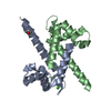
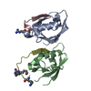
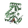
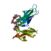
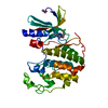
| Title | Crystal structure of calcium-free human S100Z | ||||||
 Components Components | Protein S100-Z | ||||||
 Keywords Keywords | PROTEIN BINDING / S100 / calcium-promoted oligomerisation neuronal development | ||||||
| Function / homology |  Function and homology information Function and homology informationcalcium-dependent protein binding / calcium ion binding / protein homodimerization activity Similarity search - Function | ||||||
| Biological species |  Homo sapiens (human) Homo sapiens (human) | ||||||
| Method |  X-RAY DIFFRACTION / SYNCHROTRON / MOLECULAR REPLACEMENT / Resolution: 2.3 Å X-RAY DIFFRACTION / SYNCHROTRON / MOLECULAR REPLACEMENT / Resolution: 2.3 Å | ||||||
 Authors Authors | Calderone, V. / Fragai, M. / Luchinat, C. / Gallo, G. | ||||||
 Citation Citation | Journal: J. Biol. Inorg. Chem. / Year: 2017 Title: Solving the crystal structure of human calcium-free S100Z: the siege and conquer of one of the last S100 family strongholds. Authors: Calderone, V. / Fragai, M. / Gallo, G. / Luchinat, C. #1: Journal: J. Mol. Biol. / Year: 2011Title: The crystal structure of zebrafish S100Z: implications for calcium-promoted S100 protein oligomerisation. Authors: Moroz, O.V. / Bronstein, I.B. / Wilson, K.S. | ||||||
| History |
|
- Structure visualization
| Structure viewer | Molecule: MolmilJmol/JSmol |
|---|
- Downloads & links
-Download
| PDBx/mmCIF format | 5hyd.cif.gz | 167.3 KB | Display | PDBx/mmCIF format |
|---|---|---|---|---|
| PDB format | pdb5hyd.ent.gz | 136.3 KB | Display | PDB format |
| PDBx/mmJSON format | 5hyd.json.gz | Tree view | PDBx/mmJSON format | |
| Others |  Other downloads Other downloads |
-Validation report
| Summary document | 5hyd_validation.pdf.gz | 455.6 KB | Display | wwPDB validaton report |
|---|---|---|---|---|
| Full document | 5hyd_full_validation.pdf.gz | 463.2 KB | Display | |
| Data in XML | 5hyd_validation.xml.gz | 15.6 KB | Display | |
| Data in CIF | 5hyd_validation.cif.gz | 21.1 KB | Display | |
| Arichive directory | https://data.pdbj.org/pub/pdb/validation_reports/hy/5hydftp://data.pdbj.org/pub/pdb/validation_reports/hy/5hyd | HTTPS FTP |
-Related structure data
| Related structure data | 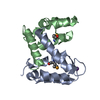 2wceS S: Starting model for refinement |
|---|---|
| Similar structure data |


-Links
 PDBj
PDBj- Assembly
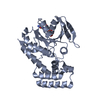
| Deposited unit | 
| ||||||||
|---|---|---|---|---|---|---|---|---|---|
| 1 | 
| ||||||||
| 2 | 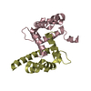
| ||||||||
| Unit cell |
|
-Components
| #1: Protein | Mass: 11637.593 Da / Num. of mol.: 4 Source method: isolated from a genetically manipulated source Source: (gene. exp.) Homo sapiens (human) / Gene: S100Z / Production host:  #2: Water | ChemComp-HOH / |  Mass: 18.015 Da / Num. of mol.: 27 / Source method: isolated from a natural source / Formula: H2O Mass: 18.015 Da / Num. of mol.: 27 / Source method: isolated from a natural source / Formula: H2OHas protein modification | Y | |
|---|
-Experimental details
-Experiment
| Experiment | Method: X-RAY DIFFRACTION / Number of used crystals: 1 |
|---|
- Sample preparation
| Crystal | Density Matthews: 2.07 Å3/Da / Density % sol: 46 % |
|---|---|
| Crystal grow | Temperature: 293 K / Method: vapor diffusion, sitting drop / pH: 7 Details: 0.1 M sodium cacodylate, 0.2 M sodium acetate, 30% PEG 8000 |
-Data collection
| Diffraction | Mean temperature: 100 K |
|---|---|
| Diffraction source | Source: SYNCHROTRON / Site: ESRF  / Beamline: ID29 / Wavelength: 0.976 Å / Beamline: ID29 / Wavelength: 0.976 Å |
| Detector | Type: DECTRIS PILATUS 6M / Detector: PIXEL / Date: Sep 15, 2013 |
| Radiation | Protocol: SINGLE WAVELENGTH / Monochromatic (M) / Laue (L): M / Scattering type: x-ray |
| Radiation wavelength | Wavelength: 0.976 Å / Relative weight: 1 |
| Reflection | Resolution: 2.3→19.8 Å / Num. obs: 17889 / % possible obs: 97.1 % / Observed criterion σ(F): 0 / Observed criterion σ(I): 0 / Redundancy: 5.5 % / Biso Wilson estimate: 34.3 Å2 / Rmerge(I) obs: 0.13 / Net I/σ(I): 3.3 |
| Reflection shell | Resolution: 2.3→2.38 Å / Redundancy: 3.7 % / Rmerge(I) obs: 0.8 / Mean I/σ(I) obs: 1 / % possible all: 90.9 |
- Processing
| Software |
| |||||||||||||||||||||||||||||||||||||||||||||||||
|---|---|---|---|---|---|---|---|---|---|---|---|---|---|---|---|---|---|---|---|---|---|---|---|---|---|---|---|---|---|---|---|---|---|---|---|---|---|---|---|---|---|---|---|---|---|---|---|---|---|---|
| Refinement | Method to determine structure: MOLECULAR REPLACEMENT Starting model: 2WCE Resolution: 2.3→19.799 Å / SU ML: 0.28 / Cross valid method: FREE R-VALUE / σ(F): 1.34 / Phase error: 27.67 / Stereochemistry target values: ML
| |||||||||||||||||||||||||||||||||||||||||||||||||
| Solvent computation | Shrinkage radii: 0.9 Å / VDW probe radii: 1.11 Å / Solvent model: FLAT BULK SOLVENT MODEL | |||||||||||||||||||||||||||||||||||||||||||||||||
| Refinement step | Cycle: LAST / Resolution: 2.3→19.799 Å
| |||||||||||||||||||||||||||||||||||||||||||||||||
| Refine LS restraints |
| |||||||||||||||||||||||||||||||||||||||||||||||||
| LS refinement shell |
| |||||||||||||||||||||||||||||||||||||||||||||||||
| Refinement TLS params. | Method: refined / Origin x: 35.7202 Å / Origin y: -33.7561 Å / Origin z: -29.3566 Å
| |||||||||||||||||||||||||||||||||||||||||||||||||
| Refinement TLS group | Selection details: all |
