Type: MAR CCD 165 mm / Detector: CCD / Date: Jul 15, 2014
Radiation
Protocol: SINGLE WAVELENGTH / Monochromatic (M) / Laue (L): M / Scattering type: x-ray
Radiation wavelength
Wavelength: 0.9792 Å / Relative weight: 1
Reflection
Resolution: 3.1→50 Å / Num. obs: 11548 / % possible obs: 99.6 % / Redundancy: 15.2 % / Net I/σ(I): 61.93
Reflection shell
Resolution: 3.1→3.15 Å / Redundancy: 16.8 % / Rmerge(I) obs: 0.557 / Mean I/σ(I) obs: 8.39 / % possible all: 100
-
Processing
Software
Name
Version
Classification
PHENIX
1.9_1692
refinement
HKL-2000
7.04
datareduction
HKL-2000
7.04
datascaling
PHASER
2.5.6
phasing
Refinement
Method to determine structure: SAD Starting model: the Se-Met derivative of this complex Resolution: 3.1→39.75 Å / SU ML: 0.46 / Cross valid method: FREE R-VALUE / σ(F): 1.33 / Phase error: 34.69 / Stereochemistry target values: ML
Rfactor
Num. reflection
% reflection
Selection details
Rfree
0.3017
537
4.76 %
random selection
Rwork
0.2594
-
-
-
obs
0.2615
11286
97.7 %
-
Solvent computation
Shrinkage radii: 0.9 Å / VDW probe radii: 1.11 Å / Solvent model: FLAT BULK SOLVENT MODEL
Refinement step
Cycle: LAST / Resolution: 3.1→39.75 Å
Protein
Nucleic acid
Ligand
Solvent
Total
Num. atoms
2510
0
0
0
2510
Refine LS restraints
Refine-ID
Type
Dev ideal
Number
X-RAY DIFFRACTION
f_bond_d
0.011
2551
X-RAY DIFFRACTION
f_angle_d
1.46
3429
X-RAY DIFFRACTION
f_dihedral_angle_d
17.719
968
X-RAY DIFFRACTION
f_chiral_restr
0.058
383
X-RAY DIFFRACTION
f_plane_restr
0.008
436
LS refinement shell
Resolution (Å)
Rfactor Rfree
Num. reflection Rfree
Rfactor Rwork
Num. reflection Rwork
Refine-ID
% reflection obs (%)
3.0999-3.4117
0.4044
133
0.2947
2681
X-RAY DIFFRACTION
100
3.4117-3.905
0.3284
136
0.2965
2541
X-RAY DIFFRACTION
95
3.905-4.9184
0.2976
147
0.2385
2678
X-RAY DIFFRACTION
98
4.9184-39.7536
0.2678
121
0.2507
2849
X-RAY DIFFRACTION
98
+
About Yorodumi
-
News
-
Feb 9, 2022. New format data for meta-information of EMDB entries
New format data for meta-information of EMDB entries
Version 3 of the EMDB header file is now the official format.
The previous official version 1.9 will be removed from the archive.
In the structure databanks used in Yorodumi, some data are registered as the other names, "COVID-19 virus" and "2019-nCoV". Here are the details of the virus and the list of structure data.
Jan 31, 2019. EMDB accession codes are about to change! (news from PDBe EMDB page)
EMDB accession codes are about to change! (news from PDBe EMDB page)
The allocation of 4 digits for EMDB accession codes will soon come to an end. Whilst these codes will remain in use, new EMDB accession codes will include an additional digit and will expand incrementally as the available range of codes is exhausted. The current 4-digit format prefixed with “EMD-” (i.e. EMD-XXXX) will advance to a 5-digit format (i.e. EMD-XXXXX), and so on. It is currently estimated that the 4-digit codes will be depleted around Spring 2019, at which point the 5-digit format will come into force.
The EM Navigator/Yorodumi systems omit the EMD- prefix.
Related info.:Q: What is EMD? / ID/Accession-code notation in Yorodumi/EM Navigator
Yorodumi is a browser for structure data from EMDB, PDB, SASBDB, etc.
This page is also the successor to EM Navigator detail page, and also detail information page/front-end page for Omokage search.
The word "yorodu" (or yorozu) is an old Japanese word meaning "ten thousand". "mi" (miru) is to see.
Related info.:EMDB / PDB / SASBDB / Comparison of 3 databanks / Yorodumi Search / Aug 31, 2016. New EM Navigator & Yorodumi / Yorodumi Papers / Jmol/JSmol / Function and homology information / Changes in new EM Navigator and Yorodumi
 Movie
Movie Controller

 Yorodumi
 Open data
 Basic information
 Components
 Keywords
 Function and homology information
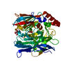
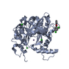
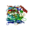
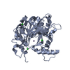
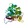
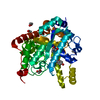
 Enterobacteria phage T4 (virus)
 X-RAY DIFFRACTION /
  Authors
 Citation
 Structure visualization
 Downloads & links
 Other downloads


 PDBj
PDBj Assembly
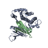
 Sample preparation
 / Beamline: 3W1A / Wavelength: 0.9792 Å
 Processing