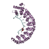
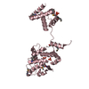
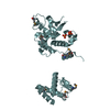
| Deposited unit | A: Tyrosine recombinase XerA
B: Tyrosine recombinase XerA
C: Tyrosine recombinase XerA
D: Tyrosine recombinase XerA
E: Tyrosine recombinase XerA
F: Tyrosine recombinase XerA
hetero molecules
| Theoretical mass | Number of molelcules |
|---|
| Total (without water) | 223,703 | 21 |
|---|
| Polymers | 222,278 | 6 |
|---|
| Non-polymers | 1,425 | 15 |
|---|
| Water | 8,845 | 491 |
|---|
|
|---|
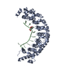
| 1 | A: Tyrosine recombinase XerA
hetero molecules
| Theoretical mass | Number of molelcules |
|---|
| Total (without water) | 37,426 | 5 |
|---|
| Polymers | 37,046 | 1 |
|---|
| Non-polymers | 380 | 4 |
|---|
| Water | 18 | 1 |
|---|
| Type | Name | Symmetry operation | Number |
|---|
| identity operation | 1_555 | x,y,z | 1 |
|
|---|
| 2 | B: Tyrosine recombinase XerA
hetero molecules
| Theoretical mass | Number of molelcules |
|---|
| Total (without water) | 37,236 | 3 |
|---|
| Polymers | 37,046 | 1 |
|---|
| Non-polymers | 190 | 2 |
|---|
| Water | 18 | 1 |
|---|
| Type | Name | Symmetry operation | Number |
|---|
| identity operation | 1_555 | x,y,z | 1 |
|
|---|
| 3 | C: Tyrosine recombinase XerA
hetero molecules
| Theoretical mass | Number of molelcules |
|---|
| Total (without water) | 37,331 | 4 |
|---|
| Polymers | 37,046 | 1 |
|---|
| Non-polymers | 285 | 3 |
|---|
| Water | 18 | 1 |
|---|
| Type | Name | Symmetry operation | Number |
|---|
| identity operation | 1_555 | x,y,z | 1 |
|
|---|
| 4 | D: Tyrosine recombinase XerA
hetero molecules
| Theoretical mass | Number of molelcules |
|---|
| Total (without water) | 37,236 | 3 |
|---|
| Polymers | 37,046 | 1 |
|---|
| Non-polymers | 190 | 2 |
|---|
| Water | 18 | 1 |
|---|
| Type | Name | Symmetry operation | Number |
|---|
| identity operation | 1_555 | x,y,z | 1 |
|
|---|
| 5 | E: Tyrosine recombinase XerA
hetero molecules
| Theoretical mass | Number of molelcules |
|---|
| Total (without water) | 37,236 | 3 |
|---|
| Polymers | 37,046 | 1 |
|---|
| Non-polymers | 190 | 2 |
|---|
| Water | 18 | 1 |
|---|
| Type | Name | Symmetry operation | Number |
|---|
| identity operation | 1_555 | x,y,z | 1 |
|
|---|
| 6 | F: Tyrosine recombinase XerA
hetero molecules
| Theoretical mass | Number of molelcules |
|---|
| Total (without water) | 37,236 | 3 |
|---|
| Polymers | 37,046 | 1 |
|---|
| Non-polymers | 190 | 2 |
|---|
| Water | 18 | 1 |
|---|
| Type | Name | Symmetry operation | Number |
|---|
| identity operation | 1_555 | x,y,z | 1 |
|
|---|
| Unit cell | | Length a, b, c (Å) | 182.860, 105.968, 115.586 |
|---|
| Angle α, β, γ (deg.) | 90.00, 110.01, 90.00 |
|---|
| Int Tables number | 5 |
|---|
| Space group name H-M | C121 |
|---|
|
|---|
 Movie
Movie Controller


 Open data
 Basic information
 Components
 Keywords
 Function and homology information
 Thermoplasma acidophilum DSM 1728 (acidophilic)
 X-RAY DIFFRACTION /
  Authors
 Korea, Republic Of, 2items
  Citation
 Structure visualization
 Downloads & links
 Other downloads


 PDBj
PDBj


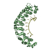
 Assembly


 Mass: 94.971 Da / Num. of mol.: 15 / Source method: obtained synthetically / Formula: PO4
 Mass: 18.015 Da / Num. of mol.: 491 / Source method: isolated from a natural source / Formula: H2O
 Sample preparation
 / Beamline: BL-5A / Wavelength: 0.97952, 0.97987, 0.9800
 Processing