 Movie
Movie Controller

[English] 日本語
 Yorodumi
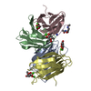
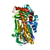
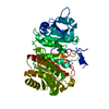
- PDB-5hv4: Crystal Structure of a Prolyl 4-Hydroxylase Complexed with Alpha-... -
+ Open data

- Basic information
| Entry | Database: PDB / ID: 5hv4 | ||||||
|---|---|---|---|---|---|---|---|
| Title | Crystal Structure of a Prolyl 4-Hydroxylase Complexed with Alpha-ketoglutarate from the Pathogenic Bacterium Bacillus anthracis in C2221 | ||||||
 Components Components | 2OG-Fe(II) oxygenase | ||||||
 Keywords Keywords | OXIDOREDUCTASE / P4H / Dioxygenase / Cupin / Fe(II)/alpha-ketoglutarate | ||||||
| Function / homology |  Function and homology information Function and homology informationprocollagen-proline 4-dioxygenase activity / L-ascorbic acid binding / iron ion binding Similarity search - Function | ||||||
| Biological species |  | ||||||
| Method |  X-RAY DIFFRACTION / SYNCHROTRON / MOLECULAR REPLACEMENT / molecular replacement / Resolution: 2.35 Å X-RAY DIFFRACTION / SYNCHROTRON / MOLECULAR REPLACEMENT / molecular replacement / Resolution: 2.35 Å | ||||||
 Authors Authors | Schnicker, N.J. / Dey, M. | ||||||
| Funding support |  United States, 1items United States, 1items
| ||||||
 Citation Citation | Journal: Acta Crystallogr D Struct Biol / Year: 2016 Title: Structural analysis of cofactor binding for a prolyl 4-hydroxylase from the pathogenic bacterium Bacillus anthracis. Authors: Schnicker, N.J. / Dey, M. | ||||||
| History |
|
- Structure visualization
| Structure viewer | Molecule: MolmilJmol/JSmol |
|---|
- Downloads & links
-Download
| PDBx/mmCIF format | 5hv4.cif.gz | 57.4 KB | Display | PDBx/mmCIF format |
|---|---|---|---|---|
| PDB format | pdb5hv4.ent.gz | 39.1 KB | Display | PDB format |
| PDBx/mmJSON format | 5hv4.json.gz | Tree view | PDBx/mmJSON format | |
| Others |  Other downloads Other downloads |
-Validation report
| Arichive directory | https://data.pdbj.org/pub/pdb/validation_reports/hv/5hv4ftp://data.pdbj.org/pub/pdb/validation_reports/hv/5hv4 | HTTPS FTP |
|---|
-Related structure data
| Related structure data |  5hv0C  3itqS C: citing same article ( S: Starting model for refinement |
|---|---|
| Similar structure data |


-Links
 PDBj
PDBj

- Assembly
| Deposited unit | 
| ||||||||
|---|---|---|---|---|---|---|---|---|---|
| 1 | 
| ||||||||
| Unit cell |
|
-Components
| #1: Protein | Mass: 24708.592 Da / Num. of mol.: 1 Source method: isolated from a genetically manipulated source Source: (gene. exp.) Gene: GBAA_4459, AB163_21210, AB164_16730, AB165_11640, AB166_03610, AB167_21505, AB168_05660, AB169_05325, AB170_09560, AB171_10145, AB893_21665, ADK17_22720, ADK18_21540, ADT20_05380, ADT21_14515, BF27_3254 Production host: | ||||||||
|---|---|---|---|---|---|---|---|---|---|
| #2: Chemical | ChemComp-CD /   Mass: 112.411 Da / Num. of mol.: 5 / Source method: obtained synthetically / Formula: Cd Mass: 112.411 Da / Num. of mol.: 5 / Source method: obtained synthetically / Formula: Cd#3: Chemical | ChemComp-AKG / |   Mass: 146.098 Da / Num. of mol.: 1 / Source method: obtained synthetically / Formula: C5H6O5 Mass: 146.098 Da / Num. of mol.: 1 / Source method: obtained synthetically / Formula: C5H6O5#4: Chemical | ChemComp-K /   Mass: 39.098 Da / Num. of mol.: 6 / Source method: obtained synthetically / Formula: K Mass: 39.098 Da / Num. of mol.: 6 / Source method: obtained synthetically / Formula: K#5: Water | ChemComp-HOH / |  Mass: 18.015 Da / Num. of mol.: 106 / Source method: isolated from a natural source / Formula: H2O Mass: 18.015 Da / Num. of mol.: 106 / Source method: isolated from a natural source / Formula: H2OSequence details | Authors state that this is a cloning artifact from the restriction site. | |
-Experimental details
-Experiment
| Experiment | Method: X-RAY DIFFRACTION / Number of used crystals: 1 |
|---|
- Sample preparation
| Crystal | Density Matthews: 2.36 Å3/Da / Density % sol: 47.94 % |
|---|---|
| Crystal grow | Temperature: 293 K / Method: vapor diffusion, hanging drop / pH: 7.8 Details: 0.1 M HEPES pH 7.8, 0.05 M Cadmium sulfate, 0.9 M Sodium acetate tri-hydrate, 0.1 M Ammonium sulfate |
-Data collection
| Diffraction | Mean temperature: 100 K |
|---|---|
| Diffraction source | Source: SYNCHROTRON / Site: ALS / Beamline: 4.2.2 / Wavelength: 1 Å |
| Detector | Type: NOIR-1 / Detector: CCD / Date: Mar 2, 2014 |
| Radiation | Protocol: SINGLE WAVELENGTH / Monochromatic (M) / Laue (L): M / Scattering type: x-ray |
| Radiation wavelength | Wavelength: 1 Å / Relative weight: 1 |
| Reflection | Resolution: 2.35→52.372 Å / Num. obs: 10150 / % possible obs: 99.9 % / Redundancy: 7 % / Biso Wilson estimate: 24.68 Å2 / Rsym value: 0.098 / Net I/σ(I): 15.8 |
| Reflection shell | Resolution: 2.35→2.48 Å / Redundancy: 7.3 % / Rmerge(I) obs: 0.377 / Mean I/σ(I) obs: 1.9 / % possible all: 99.9 |
-Phasing
| Phasing | Method: molecular replacement |
|---|
- Processing
| Software |
| |||||||||||||||||||||||||||||||||||
|---|---|---|---|---|---|---|---|---|---|---|---|---|---|---|---|---|---|---|---|---|---|---|---|---|---|---|---|---|---|---|---|---|---|---|---|---|
| Refinement | Method to determine structure: MOLECULAR REPLACEMENT Starting model: 3ITQ Resolution: 2.35→52.37 Å / SU ML: 0.28 / Cross valid method: FREE R-VALUE / σ(F): 1.34 / Phase error: 24.57 / Stereochemistry target values: ML
| |||||||||||||||||||||||||||||||||||
| Solvent computation | Shrinkage radii: 0.9 Å / VDW probe radii: 1.11 Å / Solvent model: FLAT BULK SOLVENT MODEL | |||||||||||||||||||||||||||||||||||
| Refinement step | Cycle: LAST / Resolution: 2.35→52.37 Å
| |||||||||||||||||||||||||||||||||||
| Refine LS restraints |
| |||||||||||||||||||||||||||||||||||
| LS refinement shell |
|
