 Movie
Movie Controller

[English] 日本語
 Yorodumi
- PDB-5hu6: Structure of the T. brucei haptoglobin-haemoglobin receptor bound... -
+ Open data

- Basic information
| Entry | Database: PDB / ID: 5hu6 | |||||||||
|---|---|---|---|---|---|---|---|---|---|---|
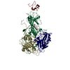
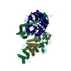
| Title | Structure of the T. brucei haptoglobin-haemoglobin receptor bound to human haptolgobin-haemoglobin | |||||||||
 Components Components |
| |||||||||
 Keywords Keywords | TRANSPORT PROTEIN / Trypanosome haptoglobin-haemoglobin | |||||||||
| Function / homology |  Function and homology information Function and homology informationciliary pocket / negative regulation of hydrogen peroxide catabolic process / heme transmembrane transporter activity / zymogen activation / cellular oxidant detoxification / Heme assimilation / nitric oxide transport / hemoglobin alpha binding / hemoglobin binding / renal absorption ...ciliary pocket / negative regulation of hydrogen peroxide catabolic process / heme transmembrane transporter activity / zymogen activation / cellular oxidant detoxification / Heme assimilation / nitric oxide transport / hemoglobin alpha binding / hemoglobin binding / renal absorption / haptoglobin-hemoglobin complex / antioxidant activity / hemoglobin complex / oxygen transport / Scavenging of heme from plasma / erythrocyte development / immune system process / endocytic vesicle lumen / receptor-mediated endocytosis / blood vessel diameter maintenance / acute-phase response / hydrogen peroxide catabolic process / oxygen carrier activity / response to hydrogen peroxide / carbon dioxide transport / Heme signaling / Erythrocytes take up oxygen and release carbon dioxide / Erythrocytes take up carbon dioxide and release oxygen / defense response / Cytoprotection by HMOX1 / oxygen binding / Late endosomal microautophagy / platelet aggregation / regulation of blood pressure / specific granule lumen / positive regulation of nitric oxide biosynthetic process / Chaperone Mediated Autophagy / tertiary granule lumen / Factors involved in megakaryocyte development and platelet production / response to oxidative stress / blood microparticle / ficolin-1-rich granule lumen / defense response to bacterium / iron ion binding / inflammatory response / serine-type endopeptidase activity / heme binding / Neutrophil degranulation / : / extracellular exosome / extracellular region / membrane / metal ion binding / cytosol Similarity search - Function | |||||||||
| Biological species |  Homo sapiens (human) Homo sapiens (human) | |||||||||
| Method |  X-RAY DIFFRACTION / SYNCHROTRON / MOLECULAR REPLACEMENT / Resolution: 2.9 Å X-RAY DIFFRACTION / SYNCHROTRON / MOLECULAR REPLACEMENT / Resolution: 2.9 Å | |||||||||
 Authors Authors | Lane-Serff, H. / Higgins, M.K. | |||||||||
 Citation Citation | Journal: Elife / Year: 2014 Title: Structural basis for ligand and innate immunity factor uptake by the trypanosome haptoglobin-haemoglobin receptor. Authors: Lane-Serff, H. / MacGregor, P. / Lowe, E.D. / Carrington, M. / Higgins, M.K. | |||||||||
| History |
|
- Structure visualization
| Structure viewer | Molecule: MolmilJmol/JSmol |
|---|
- Downloads & links
-Download
| PDBx/mmCIF format | 5hu6.cif.gz | 324.9 KB | Display | PDBx/mmCIF format |
|---|---|---|---|---|
| PDB format | pdb5hu6.ent.gz | 262.9 KB | Display | PDB format |
| PDBx/mmJSON format | 5hu6.json.gz | Tree view | PDBx/mmJSON format | |
| Others |  Other downloads Other downloads |
-Validation report
| Arichive directory | https://data.pdbj.org/pub/pdb/validation_reports/hu/5hu6ftp://data.pdbj.org/pub/pdb/validation_reports/hu/5hu6 | HTTPS FTP |
|---|
-Related structure data
| Related structure data |  4x0jC  4x0lC  4xolS S: Starting model for refinement C: citing same article ( |
|---|---|
| Similar structure data |


-Links
 PDBj
PDBj


- Assembly
| Deposited unit | 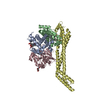
| ||||||||
|---|---|---|---|---|---|---|---|---|---|
| 1 |
| ||||||||
| Unit cell |
|
-Components
-Hemoglobin subunit ... , 2 types, 2 molecules AB
| #1: Protein | Mass: 15150.353 Da / Num. of mol.: 1 Source method: isolated from a genetically manipulated source Source: (gene. exp.) Homo sapiens (human) / Gene: HBA1, HBA2 / Production host: Homo sapiens (human) / References: UniProt: P69905 |
|---|---|
| #2: Protein | Mass: 15222.419 Da / Num. of mol.: 1 Source method: isolated from a genetically manipulated source Source: (gene. exp.) Homo sapiens (human) / Gene: HBB / Production host: Homo sapiens (human) / References: UniProt: P68871 |
-Protein , 2 types, 2 molecules CD
| #3: Protein | Mass: 28790.809 Da / Num. of mol.: 1 Source method: isolated from a genetically manipulated source Source: (gene. exp.) Homo sapiens (human) / Gene: HP / Production host:   Spodoptera frugiperda (fall armyworm) / References: UniProt: P00738 Spodoptera frugiperda (fall armyworm) / References: UniProt: P00738 |
|---|---|
| #4: Protein | Mass: 27930.453 Da / Num. of mol.: 1 Source method: isolated from a genetically manipulated source Source: (gene. exp.)  |
-Sugars , 1 types, 2 molecules 
| #7: Sugar |  Type: D-saccharide, beta linking / Mass: 221.208 Da / Num. of mol.: 2 Type: D-saccharide, beta linking / Mass: 221.208 Da / Num. of mol.: 2Source method: isolated from a genetically manipulated source Formula: C8H15NO6 |
|---|
-Non-polymers , 2 types, 4 molecules 

| #5: Chemical |  Mass: 616.487 Da / Num. of mol.: 2 / Source method: obtained synthetically / Formula: C34H32FeN4O4 Mass: 616.487 Da / Num. of mol.: 2 / Source method: obtained synthetically / Formula: C34H32FeN4O4#6: Chemical |  Mass: 31.999 Da / Num. of mol.: 2 / Source method: obtained synthetically / Formula: O2 Mass: 31.999 Da / Num. of mol.: 2 / Source method: obtained synthetically / Formula: O2 |
|---|
-Details
| Has protein modification | Y |
|---|
-Experimental details
-Experiment
| Experiment | Method: X-RAY DIFFRACTION |
|---|
- Sample preparation
| Crystal | Density Matthews: 2.43 Å3/Da / Density % sol: 49.43 % |
|---|---|
| Crystal grow | Temperature: 291 K / Method: vapor diffusion, sitting drop Details: 12.5% v/v MPD, 0.03 M NaBr, 0.03M NaI, 0.03M NaF, 0.1 M MES/imidazole pH 6.5, 12.5% w/v PEG 1000, 12.5% w/v PEG 3350 |
-Data collection
| Diffraction | Mean temperature: 100 K |
|---|---|
| Diffraction source | Source: SYNCHROTRON / Site: Diamond  / Beamline: I03 / Wavelength: 0.975 Å / Beamline: I03 / Wavelength: 0.975 Å |
| Detector | Type: DECTRIS PILATUS 6M / Detector: PIXEL / Date: Apr 21, 2014 |
| Radiation | Protocol: SINGLE WAVELENGTH / Monochromatic (M) / Laue (L): M / Scattering type: x-ray |
| Radiation wavelength | Wavelength: 0.975 Å / Relative weight: 1 |
| Reflection | Resolution: 2.9→66 Å / Num. obs: 18486 / % possible obs: 99.1 % / Redundancy: 2.7 % / Biso Wilson estimate: 76.4 Å2 / Rmerge(I) obs: 0.102 / Net I/σ(I): 6 |
| Reflection shell | Resolution: 2.9→3.06 Å / Redundancy: 2.7 % / Rmerge(I) obs: 0.42 / Mean I/σ(I) obs: 1.9 / % possible all: 98.7 |
- Processing
| Software |
| |||||||||||||||||||||||||||||||||||||||||||||||||||||||||||||||||||||||||||||||||||||||||||||||||||||||||||||||||||||||||||||
|---|---|---|---|---|---|---|---|---|---|---|---|---|---|---|---|---|---|---|---|---|---|---|---|---|---|---|---|---|---|---|---|---|---|---|---|---|---|---|---|---|---|---|---|---|---|---|---|---|---|---|---|---|---|---|---|---|---|---|---|---|---|---|---|---|---|---|---|---|---|---|---|---|---|---|---|---|---|---|---|---|---|---|---|---|---|---|---|---|---|---|---|---|---|---|---|---|---|---|---|---|---|---|---|---|---|---|---|---|---|---|---|---|---|---|---|---|---|---|---|---|---|---|---|---|---|---|
| Refinement | Method to determine structure: MOLECULAR REPLACEMENT Starting model: 4XOL Resolution: 2.9→58.07 Å / Cor.coef. Fo:Fc: 0.9219 / Cor.coef. Fo:Fc free: 0.8765 / Cross valid method: THROUGHOUT / σ(F): 0 / SU Rfree Blow DPI: 0.368
| |||||||||||||||||||||||||||||||||||||||||||||||||||||||||||||||||||||||||||||||||||||||||||||||||||||||||||||||||||||||||||||
| Displacement parameters | Biso mean: 67.95 Å2
| |||||||||||||||||||||||||||||||||||||||||||||||||||||||||||||||||||||||||||||||||||||||||||||||||||||||||||||||||||||||||||||
| Refine analyze | Luzzati coordinate error obs: 0.441 Å | |||||||||||||||||||||||||||||||||||||||||||||||||||||||||||||||||||||||||||||||||||||||||||||||||||||||||||||||||||||||||||||
| Refinement step | Cycle: 1 / Resolution: 2.9→58.07 Å
| |||||||||||||||||||||||||||||||||||||||||||||||||||||||||||||||||||||||||||||||||||||||||||||||||||||||||||||||||||||||||||||
| Refine LS restraints |
| |||||||||||||||||||||||||||||||||||||||||||||||||||||||||||||||||||||||||||||||||||||||||||||||||||||||||||||||||||||||||||||
| LS refinement shell | Resolution: 2.9→3.08 Å / Total num. of bins used: 9
| |||||||||||||||||||||||||||||||||||||||||||||||||||||||||||||||||||||||||||||||||||||||||||||||||||||||||||||||||||||||||||||
| Refinement TLS params. | Method: refined / Refine-ID: X-RAY DIFFRACTION
| |||||||||||||||||||||||||||||||||||||||||||||||||||||||||||||||||||||||||||||||||||||||||||||||||||||||||||||||||||||||||||||
| Refinement TLS group |
|
