 Movie
Movie Controller

[English] 日本語
 Yorodumi
- PDB-1dkg: CRYSTAL STRUCTURE OF THE NUCLEOTIDE EXCHANGE FACTOR GRPE BOUND TO... -
+ Open data

- Basic information
| Entry | Database: PDB / ID: 1dkg | ||||||
|---|---|---|---|---|---|---|---|
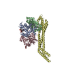
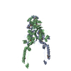
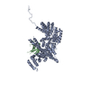
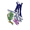
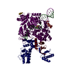
| Title | CRYSTAL STRUCTURE OF THE NUCLEOTIDE EXCHANGE FACTOR GRPE BOUND TO THE ATPASE DOMAIN OF THE MOLECULAR CHAPERONE DNAK | ||||||
 Components Components |
| ||||||
 Keywords Keywords | COMPLEX (HSP24/HSP70) / HSP70 / GRPE / MOLECULAR CHAPERONE / NUCLEOTIDE EXCHANGE FACTOR / COILED-COIL / COMPLEX (HSP24-HSP70) / COMPLEX (HSP24-HSP70) complex | ||||||
| Function / homology |  Function and homology information Function and homology information: / stress response to copper ion / sigma factor antagonist activity / adenyl-nucleotide exchange factor activity / protein-containing complex disassembly / protein unfolding / cellular response to unfolded protein / heat shock protein binding / protein folding chaperone / inclusion body ...: / stress response to copper ion / sigma factor antagonist activity / adenyl-nucleotide exchange factor activity / protein-containing complex disassembly / protein unfolding / cellular response to unfolded protein / heat shock protein binding / protein folding chaperone / inclusion body / ATP-dependent protein folding chaperone / ADP binding / : / response to heat / protein refolding / protein folding / protein-folding chaperone binding / protein-containing complex assembly / DNA replication / protein domain specific binding / protein homodimerization activity / ATP hydrolysis activity / protein-containing complex / zinc ion binding / ATP binding / membrane / plasma membrane / cytoplasm / cytosol Similarity search - Function | ||||||
| Biological species |  | ||||||
| Method |  X-RAY DIFFRACTION / SYNCHROTRON / MIR / Resolution: 2.8 Å X-RAY DIFFRACTION / SYNCHROTRON / MIR / Resolution: 2.8 Å | ||||||
 Authors Authors | Harrison, C.J. / Kuriyan, J. | ||||||
 Citation Citation | Journal: Science / Year: 1997 Title: Crystal structure of the nucleotide exchange factor GrpE bound to the ATPase domain of the molecular chaperone DnaK. Authors: Harrison, C.J. / Hayer-Hartl, M. / Di Liberto, M. / Hartl, F. / Kuriyan, J. | ||||||
| History |
|
- Structure visualization
| Structure viewer | Molecule: MolmilJmol/JSmol |
|---|
- Downloads & links
-Download
| PDBx/mmCIF format | 1dkg.cif.gz | 139.5 KB | Display | PDBx/mmCIF format |
|---|---|---|---|---|
| PDB format | pdb1dkg.ent.gz | 108.8 KB | Display | PDB format |
| PDBx/mmJSON format | 1dkg.json.gz | Tree view | PDBx/mmJSON format | |
| Others |  Other downloads Other downloads |
-Validation report
| Arichive directory | https://data.pdbj.org/pub/pdb/validation_reports/dk/1dkgftp://data.pdbj.org/pub/pdb/validation_reports/dk/1dkg | HTTPS FTP |
|---|
-Related structure data
| Similar structure data |
|---|


-Links
 PDBj
PDBj
- Assembly
| Deposited unit | 
| ||||||||
|---|---|---|---|---|---|---|---|---|---|
| 1 |
| ||||||||
| Unit cell |
|
-Components
| #1: Protein | Mass: 21882.854 Da / Num. of mol.: 2 / Mutation: CHAIN A, B, G122D Source method: isolated from a genetically manipulated source Details: ELASTASE PROTEOLYSIS PRODUCT, RESIDUES 34 - 197 / Source: (gene. exp.) #2: Protein | | Mass: 41572.105 Da / Num. of mol.: 1 / Fragment: ATPASE DOMAIN RESIDUES 3 - 383 / Mutation: CHAIN D, P319L Source method: isolated from a genetically manipulated source Source: (gene. exp.) #3: Water | ChemComp-HOH / |  Mass: 18.015 Da / Num. of mol.: 27 / Source method: isolated from a natural source / Formula: H2O Mass: 18.015 Da / Num. of mol.: 27 / Source method: isolated from a natural source / Formula: H2O |
|---|
-Experimental details
-Experiment
| Experiment | Method: X-RAY DIFFRACTION / Number of used crystals: 1 |
|---|
- Sample preparation
| Crystal | Density Matthews: 3.5 Å3/Da / Density % sol: 65 % | ||||||||||||||||||||||||||||||||||||||||
|---|---|---|---|---|---|---|---|---|---|---|---|---|---|---|---|---|---|---|---|---|---|---|---|---|---|---|---|---|---|---|---|---|---|---|---|---|---|---|---|---|---|
| Crystal grow | pH: 4.6 / Details: pH 4.6 | ||||||||||||||||||||||||||||||||||||||||
| Crystal grow | *PLUS Temperature: 4 ℃ / Method: vapor diffusion | ||||||||||||||||||||||||||||||||||||||||
| Components of the solutions | *PLUS
|
-Data collection
| Diffraction | Mean temperature: 100 K |
|---|---|
| Diffraction source | Source: SYNCHROTRON / Site: CHESS  / Beamline: F1 / Wavelength: 0.918 / Beamline: F1 / Wavelength: 0.918 |
| Detector | Detector: CCD / Date: Oct 1, 1996 |
| Radiation | Monochromatic (M) / Laue (L): M / Scattering type: x-ray |
| Radiation wavelength | Wavelength: 0.918 Å / Relative weight: 1 |
| Reflection | Resolution: 2.8→30 Å / Num. obs: 26024 / % possible obs: 95.7 % / Observed criterion σ(I): 0 / Redundancy: 29 % / Rsym value: 0.093 / Net I/σ(I): 18 |
| Reflection shell | Resolution: 2.8→2.9 Å / Mean I/σ(I) obs: 7 / Rsym value: 0.179 / % possible all: 86.4 |
| Reflection | *PLUS Num. measured all: 754420 / Rmerge(I) obs: 0.093 |
| Reflection shell | *PLUS % possible obs: 86.4 % / Rmerge(I) obs: 0.179 |
- Processing
| Software |
| ||||||||||||||||||||
|---|---|---|---|---|---|---|---|---|---|---|---|---|---|---|---|---|---|---|---|---|---|
| Refinement | Method to determine structure: MIR / Resolution: 2.8→30 Å / Rfactor Rfree error: 0.009 / Data cutoff high absF: 1000000 / Data cutoff low absF: 0.0001 / Cross valid method: THROUGHOUT / σ(F): 2 / Details: BULK SOLVENT CORRECTION, TORSION ANGLE REFINEMENT
| ||||||||||||||||||||
| Refinement step | Cycle: LAST / Resolution: 2.8→30 Å
| ||||||||||||||||||||
| Xplor file |
| ||||||||||||||||||||
| Software | *PLUS Name: X-PLOR / Version: 3.851 / Classification: refinement | ||||||||||||||||||||
| Refinement | *PLUS | ||||||||||||||||||||
| Solvent computation | *PLUS | ||||||||||||||||||||
| Displacement parameters | *PLUS | ||||||||||||||||||||
| Refine LS restraints | *PLUS
|
