[English] 日本語
Yorodumi
- PDB-5hsp: MamM CTD M250L -

+
Open data


ID or keywords:

Loading...

-
Basic information

Entry
Database: PDB / ID: 5hsp
TitleMamM CTD M250L
ComponentsMagnetosome protein MamM
KeywordsMETAL TRANSPORT / cation diffusion facilitator
Function / homology
Function and homology information


magnetosome membrane / monoatomic cation transmembrane transporter activity / iron ion transport / metal ion binding / plasma membrane
Similarity search - Function
Cation efflux protein, cytoplasmic domain / Cation efflux protein, cytoplasmic domain / Dimerisation domain of Zinc Transporter / Cation efflux protein, cytoplasmic domain superfamily / Cation efflux protein / Cation efflux transmembrane domain superfamily / Cation efflux family / Alpha-Beta Plaits / 2-Layer Sandwich / Alpha Beta
Similarity search - Domain/homology
Magnetosome protein MamM / Magnetosome protein MamM
Similarity search - Component
Biological speciesMagnetospirillum gryphiswaldense (magnetotactic)
MethodX-RAY DIFFRACTION / SYNCHROTRON / MOLECULAR REPLACEMENT / Resolution: 1.79 Å
AuthorsBarber-Zucker, S. / Zarivach, R.
Funding support Israel, 1items
OrganizationGrant numberCountry
The Israel Ministry of Science, Technology and Space Israel
CitationJournal: Sci Rep / Year: 2016
Title: Disease-Homologous Mutation in the Cation Diffusion Facilitator Protein MamM Causes Single-Domain Structural Loss and Signifies Its Importance.
Authors: Barber-Zucker, S. / Uebe, R. / Davidov, G. / Navon, Y. / Sherf, D. / Chill, J.H. / Kass, I. / Bitton, R. / Schuler, D. / Zarivach, R.
History
DepositionJan 26, 2016Deposition site: RCSB / Processing site: PDBE
Revision 1.0Nov 30, 2016Provider: repository / Type: Initial release
Revision 1.1Mar 7, 2018Group: Data collection / Category: diffrn_source / Item: _diffrn_source.pdbx_synchrotron_site
Revision 1.2Oct 16, 2019Group: Data collection / Derived calculations / Category: pdbx_struct_special_symmetry / reflns_shell
Revision 1.3Jan 10, 2024Group: Data collection / Database references / Refinement description
Category: chem_comp_atom / chem_comp_bond ...chem_comp_atom / chem_comp_bond / database_2 / pdbx_initial_refinement_model
Item: _database_2.pdbx_DOI / _database_2.pdbx_database_accession

-
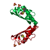
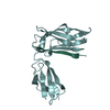
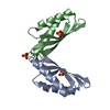
Structure visualization

Structure viewerMolecule:
MolmilJmol/JSmol

Downloads & links

-
Assembly

Deposited unit
A: Magnetosome protein MamM
D: Magnetosome protein MamM
hetero molecules


Theoretical massNumber of molelcules
Total (without water)27,7554
Polymers27,5632
Non-polymers1922
Water70339
1
A: Magnetosome protein MamM
hetero molecules

A: Magnetosome protein MamM
hetero molecules


Theoretical massNumber of molelcules
Total (without water)27,9476
Polymers27,5632
Non-polymers3844
Water362
TypeNameSymmetry operationNumber
identity operation1_555x,y,z1
crystal symmetry operation3_654-x+1,y,-z-1/21
2
D: Magnetosome protein MamM

D: Magnetosome protein MamM


Theoretical massNumber of molelcules
Total (without water)27,5632
Polymers27,5632
Non-polymers00
Water0
TypeNameSymmetry operationNumber
identity operation1_555x,y,z1
crystal symmetry operation3_654-x+1,y,-z-1/21
Unit cell
Length a, b, c (Å)37.832, 94.914, 53.685
Angle α, β, γ (deg.)90.00, 90.00, 90.00
Int Tables number20
Space group name H-MC2221
Components on special symmetry positions
IDModelComponents
11A-402-

SO4

-
Components

#1: Protein Magnetosome protein MamM / Magnetosome protein MamM / Cation efflux protein family / MamM protein


Mass: 13781.448 Da / Num. of mol.: 2 / Fragment: UNP residues 215-318
Source method: isolated from a genetically manipulated source
Source: (gene. exp.) Magnetospirillum gryphiswaldense (magnetotactic)
Gene: mamM, mgI491, MGR_4095 / Production host: Escherichia coli (E. coli) / References: UniProt: Q6NE57, UniProt: V6F235*PLUS
#2: Chemical ChemComp-SO4 / SULFATE ION


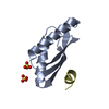
Mass: 96.063 Da / Num. of mol.: 2 / Source method: obtained synthetically / Formula: SO4
#3: Water ChemComp-HOH / water


Mass: 18.015 Da / Num. of mol.: 39 / Source method: isolated from a natural source / Formula: H2O

-
Experimental details

-
Experiment

ExperimentMethod: X-RAY DIFFRACTION / Number of used crystals: 1

-
Sample preparation

Crystal growTemperature: 293 K / Method: vapor diffusion, sitting drop
Details: 0.2 M (NH4)2SO4, 0.1 M BIS-TRIS pH=5.0 and 25% PEG 3350

-
Data collection

DiffractionMean temperature: 100 K
Diffraction sourceSource: SYNCHROTRON / Site: PETRA III, EMBL c/o DESY / Beamline: P13 (MX1) / Wavelength: 1.27 Å
DetectorType: DECTRIS PILATUS 6M / Detector: PIXEL / Date: Oct 31, 2015
RadiationProtocol: SINGLE WAVELENGTH / Monochromatic (M) / Laue (L): M / Scattering type: x-ray
Radiation wavelengthWavelength: 1.27 Å / Relative weight: 1
ReflectionResolution: 1.79→47.46 Å / Num. obs: 9313 / % possible obs: 98.7 % / Redundancy: 12.3 % / CC1/2: 0.999 / Rmerge(I) obs: 0.11 / Net I/σ(I): 16.5

-
Processing

Software
NameVersionClassification
REFMAC5.8.0135refinement
Aimlessdata scaling
PHASERphasing
XDSdata reduction
RefinementMethod to determine structure: MOLECULAR REPLACEMENT
Starting model: 3W5X
Resolution: 1.79→47.46 Å / Cor.coef. Fo:Fc: 0.963 / Cor.coef. Fo:Fc free: 0.934 / SU B: 9.032 / SU ML: 0.129 / Cross valid method: THROUGHOUT / ESU R: 0.136 / ESU R Free: 0.135 / Details: HYDROGENS HAVE BEEN ADDED IN THE RIDING POSITIONS
RfactorNum. reflection% reflectionSelection details
Rfree0.2431 978 10.5 %RANDOM
Rwork0.19687 ---
obs0.2018 8311 98.85 %-
Solvent computationIon probe radii: 0.9 Å / Shrinkage radii: 0.9 Å / VDW probe radii: 1.3 Å
Displacement parametersBiso mean: 43.794 Å2
Baniso -1Baniso -2Baniso -3
1-4.59 Å20 Å2-0 Å2
2---2.07 Å20 Å2
3----2.52 Å2
Refinement stepCycle: 1 / Resolution: 1.79→47.46 Å
ProteinNucleic acidLigandSolventTotal
Num. atoms682 0 10 39 731
Refine LS restraints
Refine-IDTypeDev idealDev ideal targetNumber
X-RAY DIFFRACTIONr_bond_refined_d0.0120.019698
X-RAY DIFFRACTIONr_bond_other_d0.0020.02676
X-RAY DIFFRACTIONr_angle_refined_deg1.4941.945944
X-RAY DIFFRACTIONr_angle_other_deg0.91431541
X-RAY DIFFRACTIONr_dihedral_angle_1_deg6.139586
X-RAY DIFFRACTIONr_dihedral_angle_2_deg37.32523.42935
X-RAY DIFFRACTIONr_dihedral_angle_3_deg14.79415119
X-RAY DIFFRACTIONr_dihedral_angle_4_deg11.138158
X-RAY DIFFRACTIONr_chiral_restr0.0870.2110
X-RAY DIFFRACTIONr_gen_planes_refined0.0050.02785
X-RAY DIFFRACTIONr_gen_planes_other0.0010.02159
X-RAY DIFFRACTIONr_nbd_refined
X-RAY DIFFRACTIONr_nbd_other
X-RAY DIFFRACTIONr_nbtor_refined
X-RAY DIFFRACTIONr_nbtor_other
X-RAY DIFFRACTIONr_xyhbond_nbd_refined
X-RAY DIFFRACTIONr_xyhbond_nbd_other
X-RAY DIFFRACTIONr_metal_ion_refined
X-RAY DIFFRACTIONr_metal_ion_other
X-RAY DIFFRACTIONr_symmetry_vdw_refined
X-RAY DIFFRACTIONr_symmetry_vdw_other
X-RAY DIFFRACTIONr_symmetry_hbond_refined
X-RAY DIFFRACTIONr_symmetry_hbond_other
X-RAY DIFFRACTIONr_symmetry_metal_ion_refined
X-RAY DIFFRACTIONr_symmetry_metal_ion_other
X-RAY DIFFRACTIONr_mcbond_it2.3572.522350
X-RAY DIFFRACTIONr_mcbond_other2.3552.51349
X-RAY DIFFRACTIONr_mcangle_it3.5013.736434
X-RAY DIFFRACTIONr_mcangle_other3.4973.75435
X-RAY DIFFRACTIONr_scbond_it3.4793.105348
X-RAY DIFFRACTIONr_scbond_other3.4573.105348
X-RAY DIFFRACTIONr_scangle_it
X-RAY DIFFRACTIONr_scangle_other5.3144.498511
X-RAY DIFFRACTIONr_long_range_B_refined8.04221.068740
X-RAY DIFFRACTIONr_long_range_B_other8.03821.073740
X-RAY DIFFRACTIONr_rigid_bond_restr
X-RAY DIFFRACTIONr_sphericity_free
X-RAY DIFFRACTIONr_sphericity_bonded
LS refinement shellResolution: 1.794→1.84 Å / Total num. of bins used: 20
RfactorNum. reflection% reflection
Rfree0.439 69 -
Rwork0.379 496 -
obs--84.33 %
Refinement TLS params.

Method: refined / Refine-ID: X-RAY DIFFRACTION

IDL112)L122)L132)L222)L232)L332)S11 (Å °)S12 (Å °)S13 (Å °)S21 (Å °)S22 (Å °)S23 (Å °)S31 (Å °)S32 (Å °)S33 (Å °)T112)T122)T132)T222)T232)T332)Origin x (Å)Origin y (Å)Origin z (Å)
12.0401-1.19090.79868.0439-4.02155.22170.0336-0.187-0.18580.20660.12320.172-0.015-0.1642-0.15680.1492-0.00560.01770.01940.01840.021318.465-14.614-0.307
27.9607-4.754-0.267514.17712.38224.34730.13830.4471-0.2108-0.44760.0807-0.34510.4692-0.0853-0.2190.25670.01220.00080.2552-0.06130.273420.968-28.433-8.51
Refinement TLS group
IDRefine-IDRefine TLS-IDAuth asym-IDAuth seq-ID
1X-RAY DIFFRACTION1A212 - 291
2X-RAY DIFFRACTION2D2 - 9

+
About Yorodumi

-
News

-
Feb 9, 2022. New format data for meta-information of EMDB entries

New format data for meta-information of EMDB entries

  • Version 3 of the EMDB header file is now the official format.
  • The previous official version 1.9 will be removed from the archive.

Related info.:EMDB header

External links:wwPDB to switch to version 3 of the EMDB data model

-
Aug 12, 2020. Covid-19 info

Covid-19 info

URL: https://pdbjlvh1.pdbj.org/emnavi/covid19.php

New page: Covid-19 featured information page in EM Navigator.

Related info.:Covid-19 info / Mar 5, 2020. Novel coronavirus structure data

+
Mar 5, 2020. Novel coronavirus structure data

Novel coronavirus structure data

Related info.:Yorodumi Speices / Aug 12, 2020. Covid-19 info

External links:COVID-19 featured content - PDBj / Molecule of the Month (242):Coronavirus Proteases

+
Jan 31, 2019. EMDB accession codes are about to change! (news from PDBe EMDB page)

EMDB accession codes are about to change! (news from PDBe EMDB page)

  • The allocation of 4 digits for EMDB accession codes will soon come to an end. Whilst these codes will remain in use, new EMDB accession codes will include an additional digit and will expand incrementally as the available range of codes is exhausted. The current 4-digit format prefixed with “EMD-” (i.e. EMD-XXXX) will advance to a 5-digit format (i.e. EMD-XXXXX), and so on. It is currently estimated that the 4-digit codes will be depleted around Spring 2019, at which point the 5-digit format will come into force.
  • The EM Navigator/Yorodumi systems omit the EMD- prefix.

Related info.:Q: What is EMD? / ID/Accession-code notation in Yorodumi/EM Navigator

External links:EMDB Accession Codes are Changing Soon! / Contact to PDBj

+
Jul 12, 2017. Major update of PDB

Major update of PDB

  • wwPDB released updated PDB data conforming to the new PDBx/mmCIF dictionary.
  • This is a major update changing the version number from 4 to 5, and with Remediation, in which all the entries are updated.
  • In this update, many items about electron microscopy experimental information are reorganized (e.g. em_software).
  • Now, EM Navigator and Yorodumi are based on the updated data.

External links:wwPDB Remediation / Enriched Model Files Conforming to OneDep Data Standards Now Available in the PDB FTP Archive

-
Yorodumi

Thousand views of thousand structures

  • Yorodumi is a browser for structure data from EMDB, PDB, SASBDB, etc.
  • This page is also the successor to EM Navigator detail page, and also detail information page/front-end page for Omokage search.
  • The word "yorodu" (or yorozu) is an old Japanese word meaning "ten thousand". "mi" (miru) is to see.

Related info.:EMDB / PDB / SASBDB / Comparison of 3 databanks / Yorodumi Search / Aug 31, 2016. New EM Navigator & Yorodumi / Yorodumi Papers / Jmol/JSmol / Function and homology information / Changes in new EM Navigator and Yorodumi

Read more