 Movie
Movie Controller

[English] 日本語
 Yorodumi
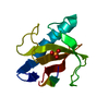
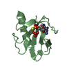
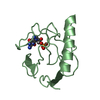
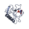
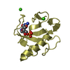
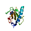
- PDB-5hoh: RIBONUCLEASE T1 (ASN9ALA/THR93ALA DOUBLEMUTANT) COMPLEXED WITH 2'GMP -
+ Open data

- Basic information
| Entry | Database: PDB / ID: 5hoh | ||||||
|---|---|---|---|---|---|---|---|
| Title | RIBONUCLEASE T1 (ASN9ALA/THR93ALA DOUBLEMUTANT) COMPLEXED WITH 2'GMP | ||||||
 Components Components | PROTEIN (RIBONUCLEASE T1) | ||||||
 Keywords Keywords | HYDROLASE / ENDORIBONUCLEASE / RIBONUCLEASE / ENDONUCLEASE | ||||||
| Function / homology |  Function and homology information Function and homology informationhyphal tip / ribonuclease T1 / ribonuclease T1 activity / cell septum / RNA endonuclease activity / lyase activity / RNA binding Similarity search - Function | ||||||
| Biological species |  | ||||||
| Method |  X-RAY DIFFRACTION / MOLECULAR REPLACEMENT / Resolution: 2 Å X-RAY DIFFRACTION / MOLECULAR REPLACEMENT / Resolution: 2 Å | ||||||
 Authors Authors | Langhorst, U. / Loris, R. / Denisov, V.P. / Doumen, J. / Roose, P. / Maes, D. / Halle, B. / Steyaert, J. | ||||||
 Citation Citation | Journal: Protein Sci. / Year: 1999 Title: Dissection of the structural and functional role of a conserved hydration site in RNase T1. Authors: Langhorst, U. / Loris, R. / Denisov, V.P. / Doumen, J. / Roose, P. / Maes, D. / Halle, B. / Steyaert, J. | ||||||
| History |
|
- Structure visualization
| Structure viewer | Molecule: MolmilJmol/JSmol |
|---|
- Downloads & links
-Download
| PDBx/mmCIF format | 5hoh.cif.gz | 96.7 KB | Display | PDBx/mmCIF format |
|---|---|---|---|---|
| PDB format | pdb5hoh.ent.gz | 74.8 KB | Display | PDB format |
| PDBx/mmJSON format | 5hoh.json.gz | Tree view | PDBx/mmJSON format | |
| Others |  Other downloads Other downloads |
-Validation report
| Summary document | 5hoh_validation.pdf.gz | 2.2 MB | Display | wwPDB validaton report |
|---|---|---|---|---|
| Full document | 5hoh_full_validation.pdf.gz | 2.2 MB | Display | |
| Data in XML | 5hoh_validation.xml.gz | 20 KB | Display | |
| Data in CIF | 5hoh_validation.cif.gz | 27.8 KB | Display | |
| Arichive directory | https://data.pdbj.org/pub/pdb/validation_reports/ho/5hohftp://data.pdbj.org/pub/pdb/validation_reports/ho/5hoh | HTTPS FTP |
-Related structure data
| Related structure data | 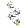 1bviC  2hohC 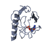 3hohC 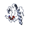 4hohC 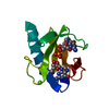 1rgaS S: Starting model for refinement C: citing same article ( |
|---|---|
| Similar structure data |


-Links
 PDBj
PDBj- Assembly
| Deposited unit | 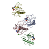
| ||||||||||||||||
|---|---|---|---|---|---|---|---|---|---|---|---|---|---|---|---|---|---|
| 1 | 
| ||||||||||||||||
| 2 | 
| ||||||||||||||||
| 3 | 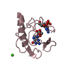
| ||||||||||||||||
| 4 | 
| ||||||||||||||||
| Unit cell |
| ||||||||||||||||
| Noncrystallographic symmetry (NCS) | NCS oper:
|
-Components
| #1: Protein | Mass: 11021.644 Da / Num. of mol.: 4 / Mutation: N9A,T93A Source method: isolated from a genetically manipulated source Details: COMPLEXED WITH GUANOSINE-2'-MONOPHOSPHATE / Source: (gene. exp.)  #2: Chemical | ChemComp-2GP /   Mass: 363.221 Da / Num. of mol.: 5 / Source method: obtained synthetically / Formula: C10H14N5O8P Mass: 363.221 Da / Num. of mol.: 5 / Source method: obtained synthetically / Formula: C10H14N5O8P#3: Chemical |   Mass: 40.078 Da / Num. of mol.: 2 / Source method: obtained synthetically / Formula: Ca Mass: 40.078 Da / Num. of mol.: 2 / Source method: obtained synthetically / Formula: Ca#4: Water | ChemComp-HOH / |  Mass: 18.015 Da / Num. of mol.: 215 / Source method: isolated from a natural source / Formula: H2O Mass: 18.015 Da / Num. of mol.: 215 / Source method: isolated from a natural source / Formula: H2OHas protein modification | Y | |
|---|
-Experimental details
-Experiment
| Experiment | Method: X-RAY DIFFRACTION / Number of used crystals: 1 |
|---|
- Sample preparation
| Crystal | Density Matthews: 2.06 Å3/Da / Density % sol: 40.27 % |
|---|---|
| Crystal grow | pH: 7.5 / Details: 25 MM NAAC PH 4.2 6.25 MM CACL2 40.0 % MPD, pH 7.5 |
| Crystal grow | *PLUS Method: otherDetails: This particular structure is not described in this paper. |
-Data collection
| Diffraction | Mean temperature: 293 K |
|---|---|
| Diffraction source | Source: ROTATING ANODE / Type: RIGAKU RU200 / Wavelength: 1.5418 |
| Detector | Type: MARRESEARCH / Detector: IMAGE PLATE / Date: Dec 1, 1997 / Details: DUAL SLITS |
| Radiation | Monochromator: GRAPHITE / Protocol: SINGLE WAVELENGTH / Monochromatic (M) / Laue (L): M / Scattering type: x-ray |
| Radiation wavelength | Wavelength: 1.5418 Å / Relative weight: 1 |
| Reflection | Resolution: 2→50 Å / Num. obs: 25314 / % possible obs: 100 % / Redundancy: 7.26 % / Rsym value: 0.108 / Net I/σ(I): 14.28 |
| Reflection shell | Resolution: 2→2.07 Å / Redundancy: 7.2 % / Mean I/σ(I) obs: 5.03 / Rsym value: 0.423 / % possible all: 100 |
- Processing
| Software |
| ||||||||||||||||||||||||||||||||||||||||||||||||||||||||||||
|---|---|---|---|---|---|---|---|---|---|---|---|---|---|---|---|---|---|---|---|---|---|---|---|---|---|---|---|---|---|---|---|---|---|---|---|---|---|---|---|---|---|---|---|---|---|---|---|---|---|---|---|---|---|---|---|---|---|---|---|---|---|
| Refinement | Method to determine structure: MOLECULAR REPLACEMENT Starting model: PDB ENTRY 1RGA Resolution: 2→50 Å / Isotropic thermal model: RESTRAINED / Cross valid method: THROUGHOUT / σ(F): 0
| ||||||||||||||||||||||||||||||||||||||||||||||||||||||||||||
| Refinement step | Cycle: LAST / Resolution: 2→50 Å
| ||||||||||||||||||||||||||||||||||||||||||||||||||||||||||||
| Refine LS restraints |
| ||||||||||||||||||||||||||||||||||||||||||||||||||||||||||||
| LS refinement shell | Resolution: 2→2.07 Å / Total num. of bins used: 8 /
| ||||||||||||||||||||||||||||||||||||||||||||||||||||||||||||
| Xplor file |
|
