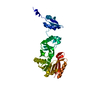
 Movie
Movie Controller


+ Open data

- Basic information
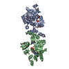
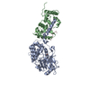
| Entry | Database: PDB / ID: 5hjm | ||||||||||||
|---|---|---|---|---|---|---|---|---|---|---|---|---|---|
| Title | Crystal Structure of Pyrococcus abyssi Trm5a complexed with MTA | ||||||||||||
 Components Components | tRNA (guanine(37)-N1)-methyltransferase Trm5a | ||||||||||||
 Keywords Keywords | TRANSFERASE / Methyltransferase / Trm5a / SAM / tRNA modification | ||||||||||||
| Function / homology |  Function and homology information Function and homology informationtRNA (guanine37-N1)-methyltransferase / tRNA (guanine(37)-N1)-methyltransferase activity / tRNA N1-guanine methylation / tRNA methylation / Transferases; Transferring one-carbon groups; Methyltransferases / cytoplasm Similarity search - Function | ||||||||||||
| Biological species |   Pyrococcus abyssi (archaea) Pyrococcus abyssi (archaea) | ||||||||||||
| Method |  X-RAY DIFFRACTION / SYNCHROTRON / MOLECULAR REPLACEMENT / Resolution: 1.762 Å X-RAY DIFFRACTION / SYNCHROTRON / MOLECULAR REPLACEMENT / Resolution: 1.762 Å | ||||||||||||
 Authors Authors | Xie, W. / Wang, C. / Jia, Q. | ||||||||||||
| Funding support |  China, 3items China, 3items
| ||||||||||||
 Citation Citation | Journal: Sci Rep / Year: 2016 Title: Crystal structures of the bifunctional tRNA methyltransferase Trm5a Authors: Wang, C. / Jia, Q. / Chen, R. / Wei, Y. / Li, J. / Ma, J. / Xie, W. | ||||||||||||
| History |
|
- Structure visualization
| Structure viewer | Molecule: MolmilJmol/JSmol |
|---|
- Downloads & links
-Download
| PDBx/mmCIF format | 5hjm.cif.gz | 98.1 KB | Display | PDBx/mmCIF format |
|---|---|---|---|---|
| PDB format | pdb5hjm.ent.gz | 71.3 KB | Display | PDB format |
| PDBx/mmJSON format | 5hjm.json.gz | Tree view | PDBx/mmJSON format | |
| Others |  Other downloads Other downloads |
-Validation report
| Arichive directory | https://data.pdbj.org/pub/pdb/validation_reports/hj/5hjmftp://data.pdbj.org/pub/pdb/validation_reports/hj/5hjm | HTTPS FTP |
|---|
-Related structure data
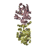
| Related structure data |  5hjiC  5hjjC  5hjkSC C: citing same article ( S: Starting model for refinement |
|---|---|
| Similar structure data |


-Links
 PDBj
PDBj- Assembly
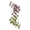
| Deposited unit | 
| ||||||||
|---|---|---|---|---|---|---|---|---|---|
| 1 |
| ||||||||
| Unit cell |
|
-Components
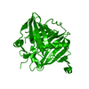
| #1: Protein | Mass: 40758.512 Da / Num. of mol.: 1 Source method: isolated from a genetically manipulated source Source: (gene. exp.) Pyrococcus abyssi (strain GE5 / Orsay) (archaea)Strain: GE5 / Orsay / Gene: trm5a, PYRAB01130, PAB2272 / Plasmid: pET-28a(+) / Production host:  References: UniProt: Q9V2G1, tRNA (guanine37-N1)-methyltransferase |
|---|---|
| #2: Chemical | ChemComp-MTA / 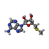  Mass: 297.334 Da / Num. of mol.: 1 Mass: 297.334 Da / Num. of mol.: 1Source method: isolated from a genetically manipulated source Formula: C11H15N5O3S |
| #3: Chemical | ChemComp-ACT /   Mass: 59.044 Da / Num. of mol.: 1 / Source method: obtained synthetically / Formula: C2H3O2 Mass: 59.044 Da / Num. of mol.: 1 / Source method: obtained synthetically / Formula: C2H3O2 |
| #4: Chemical | ChemComp-HSL /   Type: L-peptide linking / Mass: 101.104 Da / Num. of mol.: 1 / Source method: obtained synthetically / Formula: C4H7NO2 Type: L-peptide linking / Mass: 101.104 Da / Num. of mol.: 1 / Source method: obtained synthetically / Formula: C4H7NO2 |
| #5: Water | ChemComp-HOH /  Mass: 18.015 Da / Num. of mol.: 408 / Source method: isolated from a natural source / Formula: H2O Mass: 18.015 Da / Num. of mol.: 408 / Source method: isolated from a natural source / Formula: H2O |
-Experimental details
-Experiment
| Experiment | Method: X-RAY DIFFRACTION / Number of used crystals: 1 |
|---|
- Sample preparation
| Crystal | Density Matthews: 2.57 Å3/Da / Density % sol: 47.88 % |
|---|---|
| Crystal grow | Temperature: 298 K / Method: vapor diffusion, sitting drop / pH: 7.5 Details: 10-15% PEG3350, 100mM HEPES, pH7.5, 100mM Ca(OAc)2 and 100mM KCl PH range: 7.5 |
-Data collection
| Diffraction | Mean temperature: 100 K |
|---|---|
| Diffraction source | Source: SYNCHROTRON / Site: SSRF / Beamline: BL17U / Wavelength: 0.979 Å |
| Detector | Type: ADSC QUANTUM 315 / Detector: CCD / Date: Sep 27, 2015 |
| Radiation | Protocol: SINGLE WAVELENGTH / Monochromatic (M) / Laue (L): M / Scattering type: x-ray |
| Radiation wavelength | Wavelength: 0.979 Å / Relative weight: 1 |
| Reflection | Resolution: 1.76→50 Å / Num. obs: 38919 / % possible obs: 99.4 % / Observed criterion σ(I): 6.5 / Redundancy: 7 % / Rmerge(I) obs: 0.07 / Net I/σ(I): 25.1 |
| Reflection shell | Resolution: 1.76→1.82 Å / Redundancy: 7.1 % / Rmerge(I) obs: 0.324 / Mean I/σ(I) obs: 6.5 / % possible all: 95.2 |
- Processing
| Software |
| ||||||||||||||||||||||||||||||||||||||||||||||||||||||||||||||||||||||||||||||||||||||||||||||||||
|---|---|---|---|---|---|---|---|---|---|---|---|---|---|---|---|---|---|---|---|---|---|---|---|---|---|---|---|---|---|---|---|---|---|---|---|---|---|---|---|---|---|---|---|---|---|---|---|---|---|---|---|---|---|---|---|---|---|---|---|---|---|---|---|---|---|---|---|---|---|---|---|---|---|---|---|---|---|---|---|---|---|---|---|---|---|---|---|---|---|---|---|---|---|---|---|---|---|---|---|
| Refinement | Method to determine structure: MOLECULAR REPLACEMENT Starting model: 5HJK Resolution: 1.762→36.72 Å / SU ML: 0.19 / Cross valid method: FREE R-VALUE / σ(F): 1.34 / Phase error: 20.98
| ||||||||||||||||||||||||||||||||||||||||||||||||||||||||||||||||||||||||||||||||||||||||||||||||||
| Solvent computation | Shrinkage radii: 0.9 Å / VDW probe radii: 1.11 Å | ||||||||||||||||||||||||||||||||||||||||||||||||||||||||||||||||||||||||||||||||||||||||||||||||||
| Refinement step | Cycle: LAST / Resolution: 1.762→36.72 Å
| ||||||||||||||||||||||||||||||||||||||||||||||||||||||||||||||||||||||||||||||||||||||||||||||||||
| Refine LS restraints |
| ||||||||||||||||||||||||||||||||||||||||||||||||||||||||||||||||||||||||||||||||||||||||||||||||||
| LS refinement shell |
|
