 Movie
Movie Controller

[English] 日本語
 Yorodumi
- PDB-5hab: Crystal structure of mpy-RNase J (mutant H84A), an archaeal RNase... -
+ Open data

- Basic information
| Entry | Database: PDB / ID: 5hab | ||||||
|---|---|---|---|---|---|---|---|
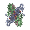
| Title | Crystal structure of mpy-RNase J (mutant H84A), an archaeal RNase J from Methanolobus psychrophilus R15, complex with RNA | ||||||
 Components Components |
| ||||||
 Keywords Keywords | HYDROLASE / exoribonuclease / beta-CASP / MBL / RNase J | ||||||
| Function / homology |  Function and homology information Function and homology information5'-3' RNA exonuclease activity / RNA catabolic process / Hydrolases; Acting on ester bonds / RNA binding / zinc ion binding / cytoplasm Similarity search - Function | ||||||
| Biological species |  Methanolobus psychrophilus R15 (archaea) Methanolobus psychrophilus R15 (archaea)synthetic construct (others) | ||||||
| Method |  X-RAY DIFFRACTION / SYNCHROTRON / MOLECULAR REPLACEMENT / Resolution: 2.3 Å X-RAY DIFFRACTION / SYNCHROTRON / MOLECULAR REPLACEMENT / Resolution: 2.3 Å | ||||||
 Authors Authors | Li, D.F. / Feng, N. | ||||||
 Citation Citation | Journal: To Be Published Title: Molecular insights into catalysis and processive exonucleolytic mechanisms of prokaryotic RNase J revealing striking parallels with that of eukaryotic Xrn1 Authors: Zheng, X. / Feng, N. / Li, D.F. / Li, J. / Dong, X.Z. | ||||||
| History |
|
- Structure visualization
| Structure viewer | Molecule: MolmilJmol/JSmol |
|---|
- Downloads & links
-Download
| PDBx/mmCIF format | 5hab.cif.gz | 201.8 KB | Display | PDBx/mmCIF format |
|---|---|---|---|---|
| PDB format | pdb5hab.ent.gz | 161.1 KB | Display | PDB format |
| PDBx/mmJSON format | 5hab.json.gz | Tree view | PDBx/mmJSON format | |
| Others |  Other downloads Other downloads |
-Validation report
| Arichive directory | https://data.pdbj.org/pub/pdb/validation_reports/ha/5habftp://data.pdbj.org/pub/pdb/validation_reports/ha/5hab | HTTPS FTP |
|---|
-Related structure data


-Links
 PDBj
PDBj

- Assembly
| Deposited unit | 
| ||||||||
|---|---|---|---|---|---|---|---|---|---|
| 1 | 
| ||||||||
| Unit cell |
| ||||||||
| Components on special symmetry positions |
|
-Components
| #1: Protein | Mass: 52113.383 Da / Num. of mol.: 2 / Fragment: UNP residues 2-448 / Mutation: H84A Source method: isolated from a genetically manipulated source Source: (gene. exp.) Methanolobus psychrophilus R15 (archaea)Gene: rnj, Mpsy_0886 / Plasmid: pET-28a / Production host:  References: UniProt: K4MAF9, Hydrolases; Acting on ester bonds #2: RNA chain | Mass: 1601.072 Da / Num. of mol.: 2 / Source method: obtained synthetically / Source: (synth.) synthetic construct (others) #3: Chemical | ChemComp-SO4 /   Mass: 96.063 Da / Num. of mol.: 9 / Source method: obtained synthetically / Formula: SO4 Mass: 96.063 Da / Num. of mol.: 9 / Source method: obtained synthetically / Formula: SO4#4: Water | ChemComp-HOH / |  Mass: 18.015 Da / Num. of mol.: 282 / Source method: isolated from a natural source / Formula: H2O Mass: 18.015 Da / Num. of mol.: 282 / Source method: isolated from a natural source / Formula: H2OHas protein modification | N | |
|---|
-Experimental details
-Experiment
| Experiment | Method: X-RAY DIFFRACTION |
|---|
- Sample preparation
| Crystal | Density Matthews: 3.29 Å3/Da / Density % sol: 62.6 % |
|---|---|
| Crystal grow | Temperature: 295 K / Method: vapor diffusion, hanging drop / pH: 7 / Details: 1.8M ammonium sulfate, 3% glycerol / PH range: 7.0-8.0 |
-Data collection
| Diffraction | Mean temperature: 100 K |
|---|---|
| Diffraction source | Source: SYNCHROTRON / Site: SSRF  / Beamline: BL19U1 / Wavelength: 0.97845 Å / Beamline: BL19U1 / Wavelength: 0.97845 Å |
| Detector | Type: PHILLIPS / Detector: PIXEL / Date: May 18, 2015 |
| Radiation | Monochromator: doubel crystal / Protocol: SINGLE WAVELENGTH / Monochromatic (M) / Laue (L): M / Scattering type: x-ray |
| Radiation wavelength | Wavelength: 0.97845 Å / Relative weight: 1 |
| Reflection | Resolution: 2.3→84.84 Å / Num. obs: 63078 / % possible obs: 99.8 % / Redundancy: 7.6 % / Biso Wilson estimate: 40 Å2 / Rmerge(I) obs: 0.105 / Rsym value: 0.105 / Net I/σ(I): 13.1 |
| Reflection shell | Resolution: 2.3→2.42 Å / Redundancy: 7.8 % / Rmerge(I) obs: 0.919 / Mean I/σ(I) obs: 2.4 / % possible all: 100 |
- Processing
| Software |
| |||||||||||||||||||||||||||||||||||||||||||||||||||||||||||||||||||||||||||||||||||||||||||||||||||||||||||||||||||||||||||||||||||||||||||||||||||||||||||||||||
|---|---|---|---|---|---|---|---|---|---|---|---|---|---|---|---|---|---|---|---|---|---|---|---|---|---|---|---|---|---|---|---|---|---|---|---|---|---|---|---|---|---|---|---|---|---|---|---|---|---|---|---|---|---|---|---|---|---|---|---|---|---|---|---|---|---|---|---|---|---|---|---|---|---|---|---|---|---|---|---|---|---|---|---|---|---|---|---|---|---|---|---|---|---|---|---|---|---|---|---|---|---|---|---|---|---|---|---|---|---|---|---|---|---|---|---|---|---|---|---|---|---|---|---|---|---|---|---|---|---|---|---|---|---|---|---|---|---|---|---|---|---|---|---|---|---|---|---|---|---|---|---|---|---|---|---|---|---|---|---|---|---|---|
| Refinement | Method to determine structure: MOLECULAR REPLACEMENT / Resolution: 2.3→55.21 Å / SU ML: 0.31 / Cross valid method: FREE R-VALUE / σ(F): 1.34 / Phase error: 26.04 / Stereochemistry target values: ML
| |||||||||||||||||||||||||||||||||||||||||||||||||||||||||||||||||||||||||||||||||||||||||||||||||||||||||||||||||||||||||||||||||||||||||||||||||||||||||||||||||
| Solvent computation | Shrinkage radii: 0.9 Å / VDW probe radii: 1.11 Å / Solvent model: FLAT BULK SOLVENT MODEL | |||||||||||||||||||||||||||||||||||||||||||||||||||||||||||||||||||||||||||||||||||||||||||||||||||||||||||||||||||||||||||||||||||||||||||||||||||||||||||||||||
| Displacement parameters | Biso mean: 50.2 Å2 | |||||||||||||||||||||||||||||||||||||||||||||||||||||||||||||||||||||||||||||||||||||||||||||||||||||||||||||||||||||||||||||||||||||||||||||||||||||||||||||||||
| Refinement step | Cycle: LAST / Resolution: 2.3→55.21 Å
| |||||||||||||||||||||||||||||||||||||||||||||||||||||||||||||||||||||||||||||||||||||||||||||||||||||||||||||||||||||||||||||||||||||||||||||||||||||||||||||||||
| Refine LS restraints |
| |||||||||||||||||||||||||||||||||||||||||||||||||||||||||||||||||||||||||||||||||||||||||||||||||||||||||||||||||||||||||||||||||||||||||||||||||||||||||||||||||
| LS refinement shell |
|
