[English] 日本語
Yorodumi
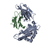
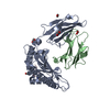
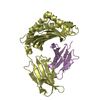
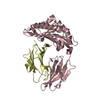
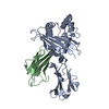
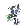
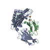
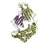
- PDB-5h94: Crystal structure of Swine MHC CLASSI for 1.48 angstroms -

+
Open data


ID or keywords:

Loading...

-
Basic information

Entry
Database: PDB / ID: 5h94
TitleCrystal structure of Swine MHC CLASSI for 1.48 angstroms
Components
  • Beta-2-microglobulin
  • MHC class I antigen
  • Nonapeptide from Influenza A virus HA protein
KeywordsIMMUNE SYSTEM / Swine / Crystal structure / MHC I / Influenza A virus / Mechanism / CTL immunity
Function / homology
Function and homology information


ER-Phagosome pathway / Endosomal/Vacuolar pathway / DAP12 interactions / DAP12 signaling / Antigen Presentation: Folding, assembly and peptide loading of class I MHC / Immunoregulatory interactions between a Lymphoid and a non-Lymphoid cell / Neutrophil degranulation / antigen processing and presentation of peptide antigen via MHC class I / antigen processing and presentation / viral budding from plasma membrane ...ER-Phagosome pathway / Endosomal/Vacuolar pathway / DAP12 interactions / DAP12 signaling / Antigen Presentation: Folding, assembly and peptide loading of class I MHC / Immunoregulatory interactions between a Lymphoid and a non-Lymphoid cell / Neutrophil degranulation / antigen processing and presentation of peptide antigen via MHC class I / antigen processing and presentation / viral budding from plasma membrane / MHC class I protein complex / peptide antigen assembly with MHC class II protein complex / MHC class II protein complex / peptide antigen binding / antigen processing and presentation of exogenous peptide antigen via MHC class II / positive regulation of immune response / positive regulation of T cell activation / MHC class II protein complex binding / late endosome membrane / clathrin-dependent endocytosis of virus by host cell / membrane => GO:0016020 / host cell surface receptor binding / immune response / apical plasma membrane / lysosomal membrane / fusion of virus membrane with host plasma membrane / fusion of virus membrane with host endosome membrane / viral envelope / virion attachment to host cell / host cell plasma membrane / virion membrane / extracellular region
Similarity search - Function
Haemagglutinin, influenzavirus A / Haemagglutinin, HA1 chain, alpha/beta domain superfamily / Haemagglutinin / Haemagglutinin, influenzavirus A/B / Viral capsid/haemagglutinin protein / MHC class I, alpha chain, C-terminal / MHC_I C-terminus / MHC class I-like antigen recognition-like / Murine Class I Major Histocompatibility Complex, H2-DB; Chain A, domain 1 / MHC class I alpha chain, alpha1 alpha2 domains ...Haemagglutinin, influenzavirus A / Haemagglutinin, HA1 chain, alpha/beta domain superfamily / Haemagglutinin / Haemagglutinin, influenzavirus A/B / Viral capsid/haemagglutinin protein / MHC class I, alpha chain, C-terminal / MHC_I C-terminus / MHC class I-like antigen recognition-like / Murine Class I Major Histocompatibility Complex, H2-DB; Chain A, domain 1 / MHC class I alpha chain, alpha1 alpha2 domains / Class I Histocompatibility antigen, domains alpha 1 and 2 / Beta-2-Microglobulin / MHC class I-like antigen recognition-like / MHC class I-like antigen recognition-like superfamily / MHC classes I/II-like antigen recognition protein / Immunoglobulin/major histocompatibility complex, conserved site / Immunoglobulins and major histocompatibility complex proteins signature. / Immunoglobulin C-Type / Immunoglobulin C1-set / Immunoglobulin C1-set domain / Ig-like domain profile. / Immunoglobulin-like domain / Immunoglobulin-like domain superfamily / Immunoglobulins / Immunoglobulin-like fold / Immunoglobulin-like / Sandwich / 2-Layer Sandwich / Mainly Beta / Alpha Beta
Similarity search - Domain/homology
MHC class I antigen / Hemagglutinin / Beta-2-microglobulin
Similarity search - Component
Biological speciesSus scrofa (pig)
H1N1 swine influenza virus
MethodX-RAY DIFFRACTION / MOLECULAR REPLACEMENT / Resolution: 1.48 Å
AuthorsFan, S. / Zhang, N. / Wang, S. / Wu, Y. / Xia, C.
Funding support China, 1items
OrganizationGrant numberCountry
The 863 Project of the China Ministry of Science and TechnologyNo. 2013AA102503 China
CitationJournal: J.Virol. / Year: 2016
Title: Structural and Biochemical Analyses of Swine Major Histocompatibility Complex Class I Complexes and Prediction of the Epitope Map of Important Influenza A Virus Strains
Authors: Fan, S. / Wu, Y. / Wang, S. / Wang, Z. / Jiang, B. / Liu, Y. / Liang, R. / Zhou, W. / Zhang, N. / Xia, C.
History
DepositionDec 25, 2015Deposition site: RCSB / Processing site: PDBJ
Revision 1.0May 18, 2016Provider: repository / Type: Initial release
Revision 1.1May 25, 2016Group: Database references
Revision 1.2Jul 27, 2016Group: Database references
Revision 1.3Oct 4, 2017Group: Data collection / Database references / Derived calculations
Category: citation / diffrn_detector / pdbx_struct_oper_list
Item: _citation.journal_id_CSD / _diffrn_detector.detector / _pdbx_struct_oper_list.symmetry_operation
Revision 1.4Nov 8, 2023Group: Data collection / Database references / Refinement description
Category: chem_comp_atom / chem_comp_bond ...chem_comp_atom / chem_comp_bond / database_2 / pdbx_initial_refinement_model
Item: _database_2.pdbx_DOI / _database_2.pdbx_database_accession

-
Structure visualization

Structure viewerMolecule:
MolmilJmol/JSmol

Downloads & links

-
Assembly

Deposited unit
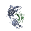
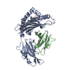
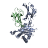
A: MHC class I antigen
B: Beta-2-microglobulin
C: Nonapeptide from Influenza A virus HA protein
D: MHC class I antigen
E: Beta-2-microglobulin
F: Nonapeptide from Influenza A virus HA protein


Theoretical massNumber of molelcules
Total (without water)88,3326
Polymers88,3326
Non-polymers00
Water14,916828
1
A: MHC class I antigen
B: Beta-2-microglobulin
C: Nonapeptide from Influenza A virus HA protein


Theoretical massNumber of molelcules
Total (without water)44,1663
Polymers44,1663
Non-polymers00
Water543
TypeNameSymmetry operationNumber
identity operation1_555x,y,z1
Buried area4300 Å2
ΔGint-18 kcal/mol
Surface area18610 Å2
MethodPISA
2
D: MHC class I antigen
E: Beta-2-microglobulin
F: Nonapeptide from Influenza A virus HA protein


Theoretical massNumber of molelcules
Total (without water)44,1663
Polymers44,1663
Non-polymers00
Water543
TypeNameSymmetry operationNumber
identity operation1_555x,y,z1
Buried area4160 Å2
ΔGint-17 kcal/mol
Surface area18630 Å2
MethodPISA
Unit cell
Length a, b, c (Å)206.453, 41.471, 106.735
Angle α, β, γ (deg.)90.00, 117.46, 90.00
Int Tables number5
Space group name H-MC121

-
Components

#1: Protein MHC class I antigen


Mass: 31693.668 Da / Num. of mol.: 2
Source method: isolated from a genetically manipulated source
Source: (gene. exp.) Sus scrofa (pig) / Gene: SLA-3*hs0202 / Production host: Escherichia coli (E. coli) / References: UniProt: A0A0N9JT91*PLUS
#2: Protein Beta-2-microglobulin / Lactollin


Mass: 11431.918 Da / Num. of mol.: 2
Source method: isolated from a genetically manipulated source
Source: (gene. exp.) Sus scrofa (pig) / Gene: B2M / Production host: Escherichia coli (E. coli) / References: UniProt: Q07717
#3: Protein/peptide Nonapeptide from Influenza A virus HA protein


Mass: 1040.212 Da / Num. of mol.: 2 / Source method: obtained synthetically / Source: (synth.) H1N1 swine influenza virus / References: UniProt: C4RUF0*PLUS
#4: Water ChemComp-HOH / water


Mass: 18.015 Da / Num. of mol.: 828 / Source method: isolated from a natural source / Formula: H2O
Sequence detailsSEQUENCE OF MHC CLASS I ANTIGEN USED IN THIS STRUCTURE HAS BEEN DEPOSITED TO DATABASE.

-
Experimental details

-
Experiment

ExperimentMethod: X-RAY DIFFRACTION / Number of used crystals: 1

-
Sample preparation

CrystalDensity Matthews: 2.3 Å3/Da / Density % sol: 46.64 %
Crystal growTemperature: 291 K / Method: vapor diffusion, hanging drop
Details: 20%(w/v) Polyethylene glycol 3350, 0.2M Ammonium fluoride

-
Data collection

DiffractionMean temperature: 100 K
Diffraction sourceSource: ROTATING ANODE / Type: RIGAKU MICROMAX-007 HF / Wavelength: 0.97892 Å
DetectorType: ADSC QUANTUM 315 / Detector: CCD / Date: Apr 30, 2014
RadiationMonochromator: NI FILTER / Protocol: SINGLE WAVELENGTH / Monochromatic (M) / Laue (L): M / Scattering type: x-ray
Radiation wavelengthWavelength: 0.97892 Å / Relative weight: 1
ReflectionResolution: 1.48→50 Å / Num. obs: 127299 / % possible obs: 99.8 % / Redundancy: 3.7 % / Net I/σ(I): 19.482
Reflection shellResolution: 1.48→1.518 Å / Redundancy: 3.6 % / Rmerge(I) obs: 0.384 / Mean I/σ(I) obs: 3.536 / % possible all: 99.8

-
Processing

Software
NameVersionClassification
REFMAC5.8.0073refinement
HKL-2000data reduction
PHASERdata scaling
PHENIXphasing
RefinementMethod to determine structure: MOLECULAR REPLACEMENT
Starting model: 3QQ4
Resolution: 1.48→50 Å / Cor.coef. Fo:Fc: 0.972 / Cor.coef. Fo:Fc free: 0.956 / SU B: 3.639 / SU ML: 0.058 / Cross valid method: THROUGHOUT / ESU R: 0.075 / ESU R Free: 0.068 / Stereochemistry target values: MAXIMUM LIKELIHOOD
RfactorNum. reflection% reflectionSelection details
Rfree0.19025 6668 5 %RANDOM
Rwork0.14975 ---
obs0.15177 127299 99.61 %-
Solvent computationIon probe radii: 0.8 Å / Shrinkage radii: 0.8 Å / VDW probe radii: 1.2 Å / Solvent model: MASK
Displacement parametersBiso mean: 21.929 Å2
Baniso -1Baniso -2Baniso -3
1-0.5 Å2-0 Å21.28 Å2
2---4.47 Å20 Å2
3---1.71 Å2
Refinement stepCycle: LAST / Resolution: 1.48→50 Å
ProteinNucleic acidLigandSolventTotal
Num. atoms6220 0 0 828 7048
Refine LS restraints
Refine-IDTypeDev idealDev ideal targetNumber
X-RAY DIFFRACTIONr_bond_refined_d0.0250.0196432
X-RAY DIFFRACTIONr_bond_other_d
X-RAY DIFFRACTIONr_angle_refined_deg2.1711.9328746
X-RAY DIFFRACTIONr_angle_other_deg
X-RAY DIFFRACTIONr_dihedral_angle_1_deg6.3535759
X-RAY DIFFRACTIONr_dihedral_angle_2_deg34.91924.062352
X-RAY DIFFRACTIONr_dihedral_angle_3_deg13.035151022
X-RAY DIFFRACTIONr_dihedral_angle_4_deg20.3941546
X-RAY DIFFRACTIONr_chiral_restr0.170.2882
X-RAY DIFFRACTIONr_gen_planes_refined0.0130.0215117
X-RAY DIFFRACTIONr_gen_planes_other
X-RAY DIFFRACTIONr_nbd_refined
X-RAY DIFFRACTIONr_nbd_other
X-RAY DIFFRACTIONr_nbtor_refined
X-RAY DIFFRACTIONr_nbtor_other
X-RAY DIFFRACTIONr_xyhbond_nbd_refined
X-RAY DIFFRACTIONr_xyhbond_nbd_other
X-RAY DIFFRACTIONr_metal_ion_refined
X-RAY DIFFRACTIONr_metal_ion_other
X-RAY DIFFRACTIONr_symmetry_vdw_refined
X-RAY DIFFRACTIONr_symmetry_vdw_other
X-RAY DIFFRACTIONr_symmetry_hbond_refined
X-RAY DIFFRACTIONr_symmetry_hbond_other
X-RAY DIFFRACTIONr_symmetry_metal_ion_refined
X-RAY DIFFRACTIONr_symmetry_metal_ion_other
X-RAY DIFFRACTIONr_mcbond_it3.2641.7883054
X-RAY DIFFRACTIONr_mcbond_other
X-RAY DIFFRACTIONr_mcangle_it3.962.7053807
X-RAY DIFFRACTIONr_mcangle_other
X-RAY DIFFRACTIONr_scbond_it5.1072.143376
X-RAY DIFFRACTIONr_scbond_other
X-RAY DIFFRACTIONr_scangle_it
X-RAY DIFFRACTIONr_scangle_other
X-RAY DIFFRACTIONr_long_range_B_refined5.17117.1910727
X-RAY DIFFRACTIONr_long_range_B_other
X-RAY DIFFRACTIONr_rigid_bond_restr8.27336430
X-RAY DIFFRACTIONr_sphericity_free28.645218
X-RAY DIFFRACTIONr_sphericity_bonded24.73356857
LS refinement shellResolution: 1.48→1.518 Å / Total num. of bins used: 20
RfactorNum. reflection% reflection
Rfree0.297 459 -
Rwork0.26 9089 -
obs--97.2 %

+
About Yorodumi

-
News

-
Feb 9, 2022. New format data for meta-information of EMDB entries

New format data for meta-information of EMDB entries

  • Version 3 of the EMDB header file is now the official format.
  • The previous official version 1.9 will be removed from the archive.

Related info.:EMDB header

External links:wwPDB to switch to version 3 of the EMDB data model

-
Aug 12, 2020. Covid-19 info

Covid-19 info

URL: https://pdbjlvh1.pdbj.org/emnavi/covid19.php

New page: Covid-19 featured information page in EM Navigator.

Related info.:Covid-19 info / Mar 5, 2020. Novel coronavirus structure data

+
Mar 5, 2020. Novel coronavirus structure data

Novel coronavirus structure data

Related info.:Yorodumi Speices / Aug 12, 2020. Covid-19 info

External links:COVID-19 featured content - PDBj / Molecule of the Month (242):Coronavirus Proteases

+
Jan 31, 2019. EMDB accession codes are about to change! (news from PDBe EMDB page)

EMDB accession codes are about to change! (news from PDBe EMDB page)

  • The allocation of 4 digits for EMDB accession codes will soon come to an end. Whilst these codes will remain in use, new EMDB accession codes will include an additional digit and will expand incrementally as the available range of codes is exhausted. The current 4-digit format prefixed with “EMD-” (i.e. EMD-XXXX) will advance to a 5-digit format (i.e. EMD-XXXXX), and so on. It is currently estimated that the 4-digit codes will be depleted around Spring 2019, at which point the 5-digit format will come into force.
  • The EM Navigator/Yorodumi systems omit the EMD- prefix.

Related info.:Q: What is EMD? / ID/Accession-code notation in Yorodumi/EM Navigator

External links:EMDB Accession Codes are Changing Soon! / Contact to PDBj

+
Jul 12, 2017. Major update of PDB

Major update of PDB

  • wwPDB released updated PDB data conforming to the new PDBx/mmCIF dictionary.
  • This is a major update changing the version number from 4 to 5, and with Remediation, in which all the entries are updated.
  • In this update, many items about electron microscopy experimental information are reorganized (e.g. em_software).
  • Now, EM Navigator and Yorodumi are based on the updated data.

External links:wwPDB Remediation / Enriched Model Files Conforming to OneDep Data Standards Now Available in the PDB FTP Archive

-
Yorodumi

Thousand views of thousand structures

  • Yorodumi is a browser for structure data from EMDB, PDB, SASBDB, etc.
  • This page is also the successor to EM Navigator detail page, and also detail information page/front-end page for Omokage search.
  • The word "yorodu" (or yorozu) is an old Japanese word meaning "ten thousand". "mi" (miru) is to see.

Related info.:EMDB / PDB / SASBDB / Comparison of 3 databanks / Yorodumi Search / Aug 31, 2016. New EM Navigator & Yorodumi / Yorodumi Papers / Jmol/JSmol / Function and homology information / Changes in new EM Navigator and Yorodumi

Read more