[English] 日本語
Yorodumi
- PDB-5h45: Crystal structure of the C-terminal Lon protease-like domain of T... -

+
Open data


ID or keywords:

Loading...

-
Basic information

Entry
Database: PDB / ID: 5h45
Title
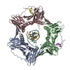
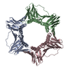
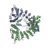
Crystal structure of the C-terminal Lon protease-like domain of Thermus thermophilus RadA/Sms
ComponentsDNA repair protein RadA
KeywordsDNA BINDING PROTEIN / Ribosomal protein S5 domain 2-like fold
Function / homology
Function and homology information


recombinational repair / ATP-dependent activity, acting on DNA / damaged DNA binding / hydrolase activity / ATP binding / metal ion binding
Similarity search - Function
DNA repair protein RadA / Subunit ChlI of Mg-chelatase / LapB, rubredoxin metal binding domain / Rubredoxin metal binding domain / DNA recombination and repair protein RecA-like, ATP-binding domain / RecA family profile 1. / Ribosomal protein S5 domain 2-type fold, subgroup / Ribosomal protein S5 domain 2-type fold / ATPases associated with a variety of cellular activities / AAA+ ATPase domain / P-loop containing nucleoside triphosphate hydrolase
Similarity search - Domain/homology
DNA repair protein RadA
Similarity search - Component
Biological speciesThermus thermophilus (bacteria)
MethodX-RAY DIFFRACTION / SYNCHROTRON / MOLECULAR REPLACEMENT / Resolution: 2.7 Å
AuthorsInoue, M. / Fukui, K. / Fujii, Y. / Nakagawa, N. / Yano, T. / Kuramitsu, S. / Masui, R.
CitationJournal: J. Biol. Chem. / Year: 2017
Title: The Lon protease-like domain in the bacterial RecA paralog RadA is required for DNA binding and repair.
Authors: Inoue, M. / Fukui, K. / Fujii, Y. / Nakagawa, N. / Yano, T. / Kuramitsu, S. / Masui, R.
History
DepositionOct 30, 2016Deposition site: PDBJ / Processing site: PDBJ
Revision 1.0May 3, 2017Provider: repository / Type: Initial release
Revision 1.1Jun 21, 2017Group: Database references / Category: citation
Item: _citation.country / _citation.journal_abbrev ..._citation.country / _citation.journal_abbrev / _citation.journal_id_ASTM / _citation.journal_id_CSD / _citation.journal_volume / _citation.page_first / _citation.page_last / _citation.pdbx_database_id_PubMed / _citation.title
Revision 1.2Mar 20, 2024Group: Data collection / Database references / Category: chem_comp_atom / chem_comp_bond / database_2
Item: _database_2.pdbx_DOI / _database_2.pdbx_database_accession

-
Structure visualization

Structure viewerMolecule:
MolmilJmol/JSmol

Downloads & links

-
Assembly

Deposited unit
A: DNA repair protein RadA
B: DNA repair protein RadA


Theoretical massNumber of molelcules
Total (without water)39,2672
Polymers39,2672
Non-polymers00
Water93752
1
A: DNA repair protein RadA
B: DNA repair protein RadA

A: DNA repair protein RadA
B: DNA repair protein RadA

A: DNA repair protein RadA
B: DNA repair protein RadA


Theoretical massNumber of molelcules
Total (without water)117,8026
Polymers117,8026
Non-polymers00
Water1086
TypeNameSymmetry operationNumber
identity operation1_555x,y,z1
crystal symmetry operation5_555z,x,y1
crystal symmetry operation9_555y,z,x1
Buried area10020 Å2
ΔGint-36 kcal/mol
Surface area36420 Å2
MethodPISA
Unit cell
Length a, b, c (Å)156.289, 156.289, 156.289
Angle α, β, γ (deg.)90.00, 90.00, 90.00
Int Tables number212
Space group name H-MP4332

-
Components

#1: Protein DNA repair protein RadA


Mass: 19633.619 Da / Num. of mol.: 2 / Fragment: UNP residues 262-423
Source method: isolated from a genetically manipulated source
Source: (gene. exp.) Thermus thermophilus (strain HB8 / ATCC 27634 / DSM 579) (bacteria)
Strain: HB8 / ATCC 27634 / DSM 579 / Gene: radA, TTHA0541 / Plasmid: pET-HisTEV / Production host: Escherichia coli (E. coli) / Strain (production host): Rosetta 2(DE3) / References: UniProt: Q5SKV2
#2: Water ChemComp-HOH / water


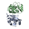
Mass: 18.015 Da / Num. of mol.: 52 / Source method: isolated from a natural source / Formula: H2O

-
Experimental details

-
Experiment

ExperimentMethod: X-RAY DIFFRACTION / Number of used crystals: 1

-
Sample preparation

CrystalDensity Matthews: 4.05 Å3/Da / Density % sol: 69.64 %
Crystal growTemperature: 277.15 K / Method: batch mode / pH: 7.5
Details: 4mg/mL protein, 50mM Tris-HCl, 500mM sodium chloride

-
Data collection

DiffractionMean temperature: 100 K
Diffraction sourceSource: SYNCHROTRON / Site: SPring-8 / Beamline: BL38B1 / Wavelength: 1 Å
DetectorType: RAYONIX MX225HE / Detector: CCD / Date: Feb 8, 2015
RadiationProtocol: SINGLE WAVELENGTH / Monochromatic (M) / Laue (L): M / Scattering type: x-ray
Radiation wavelengthWavelength: 1 Å / Relative weight: 1
ReflectionResolution: 2.7→50 Å / Num. obs: 18550 / % possible obs: 100 % / Redundancy: 41.3 % / Net I/σ(I): 20.4
Reflection shellResolution: 2.7→2.75 Å / Redundancy: 41.6 % / Mean I/σ(I) obs: 1.9 / CC1/2: 0.625 / % possible all: 100

-
Processing

Software
NameVersionClassification
REFMAC5.6.0117refinement
HKL-2000data reduction
HKL-2000data scaling
MOLREPphasing
RefinementMethod to determine structure: MOLECULAR REPLACEMENT / Resolution: 2.7→49.42 Å / Cor.coef. Fo:Fc: 0.948 / Cor.coef. Fo:Fc free: 0.911 / SU B: 8.581 / SU ML: 0.176 / Cross valid method: THROUGHOUT / ESU R: 0.289 / ESU R Free: 0.251 / Details: HYDROGENS HAVE BEEN USED IF PRESENT IN THE INPUT
RfactorNum. reflection% reflectionSelection details
Rfree0.24997 947 5.1 %RANDOM
Rwork0.19648 ---
obs0.19908 17548 99.96 %-
Solvent computationIon probe radii: 0.8 Å / Shrinkage radii: 0.8 Å / VDW probe radii: 1.2 Å
Displacement parametersBiso mean: 53.493 Å2
Refinement stepCycle: 1 / Resolution: 2.7→49.42 Å
ProteinNucleic acidLigandSolventTotal
Num. atoms2288 0 0 52 2340
Refine LS restraints
Refine-IDTypeDev idealDev ideal targetNumber
X-RAY DIFFRACTIONr_bond_refined_d0.0130.0192319
X-RAY DIFFRACTIONr_bond_other_d
X-RAY DIFFRACTIONr_angle_refined_deg1.6222.0173148
X-RAY DIFFRACTIONr_angle_other_deg
X-RAY DIFFRACTIONr_dihedral_angle_1_deg6.8695307
X-RAY DIFFRACTIONr_dihedral_angle_2_deg29.48121.48994
X-RAY DIFFRACTIONr_dihedral_angle_3_deg19.02215375
X-RAY DIFFRACTIONr_dihedral_angle_4_deg17.5061533
X-RAY DIFFRACTIONr_chiral_restr0.0940.2372
X-RAY DIFFRACTIONr_gen_planes_refined0.0080.0211759
X-RAY DIFFRACTIONr_gen_planes_other
X-RAY DIFFRACTIONr_nbd_refined
X-RAY DIFFRACTIONr_nbd_other
X-RAY DIFFRACTIONr_nbtor_refined
X-RAY DIFFRACTIONr_nbtor_other
X-RAY DIFFRACTIONr_xyhbond_nbd_refined
X-RAY DIFFRACTIONr_xyhbond_nbd_other
X-RAY DIFFRACTIONr_metal_ion_refined
X-RAY DIFFRACTIONr_metal_ion_other
X-RAY DIFFRACTIONr_symmetry_vdw_refined
X-RAY DIFFRACTIONr_symmetry_vdw_other
X-RAY DIFFRACTIONr_symmetry_hbond_refined
X-RAY DIFFRACTIONr_symmetry_hbond_other
X-RAY DIFFRACTIONr_symmetry_metal_ion_refined
X-RAY DIFFRACTIONr_symmetry_metal_ion_other
X-RAY DIFFRACTIONr_mcbond_it
X-RAY DIFFRACTIONr_mcbond_other
X-RAY DIFFRACTIONr_mcangle_it
X-RAY DIFFRACTIONr_mcangle_other
X-RAY DIFFRACTIONr_scbond_it
X-RAY DIFFRACTIONr_scbond_other
X-RAY DIFFRACTIONr_scangle_it
X-RAY DIFFRACTIONr_scangle_other
X-RAY DIFFRACTIONr_long_range_B_refined
X-RAY DIFFRACTIONr_long_range_B_other
X-RAY DIFFRACTIONr_rigid_bond_restr
X-RAY DIFFRACTIONr_sphericity_free
X-RAY DIFFRACTIONr_sphericity_bonded
LS refinement shellResolution: 2.7→2.77 Å / Total num. of bins used: 20
RfactorNum. reflection% reflection
Rfree0.343 76 -
Rwork0.267 1109 -
obs--100 %

+
About Yorodumi

-
News

-
Feb 9, 2022. New format data for meta-information of EMDB entries

New format data for meta-information of EMDB entries

  • Version 3 of the EMDB header file is now the official format.
  • The previous official version 1.9 will be removed from the archive.

Related info.:EMDB header

External links:wwPDB to switch to version 3 of the EMDB data model

-
Aug 12, 2020. Covid-19 info

Covid-19 info

URL: https://pdbjlvh1.pdbj.org/emnavi/covid19.php

New page: Covid-19 featured information page in EM Navigator.

Related info.:Covid-19 info / Mar 5, 2020. Novel coronavirus structure data

+
Mar 5, 2020. Novel coronavirus structure data

Novel coronavirus structure data

Related info.:Yorodumi Speices / Aug 12, 2020. Covid-19 info

External links:COVID-19 featured content - PDBj / Molecule of the Month (242):Coronavirus Proteases

+
Jan 31, 2019. EMDB accession codes are about to change! (news from PDBe EMDB page)

EMDB accession codes are about to change! (news from PDBe EMDB page)

  • The allocation of 4 digits for EMDB accession codes will soon come to an end. Whilst these codes will remain in use, new EMDB accession codes will include an additional digit and will expand incrementally as the available range of codes is exhausted. The current 4-digit format prefixed with “EMD-” (i.e. EMD-XXXX) will advance to a 5-digit format (i.e. EMD-XXXXX), and so on. It is currently estimated that the 4-digit codes will be depleted around Spring 2019, at which point the 5-digit format will come into force.
  • The EM Navigator/Yorodumi systems omit the EMD- prefix.

Related info.:Q: What is EMD? / ID/Accession-code notation in Yorodumi/EM Navigator

External links:EMDB Accession Codes are Changing Soon! / Contact to PDBj

+
Jul 12, 2017. Major update of PDB

Major update of PDB

  • wwPDB released updated PDB data conforming to the new PDBx/mmCIF dictionary.
  • This is a major update changing the version number from 4 to 5, and with Remediation, in which all the entries are updated.
  • In this update, many items about electron microscopy experimental information are reorganized (e.g. em_software).
  • Now, EM Navigator and Yorodumi are based on the updated data.

External links:wwPDB Remediation / Enriched Model Files Conforming to OneDep Data Standards Now Available in the PDB FTP Archive

-
Yorodumi

Thousand views of thousand structures

  • Yorodumi is a browser for structure data from EMDB, PDB, SASBDB, etc.
  • This page is also the successor to EM Navigator detail page, and also detail information page/front-end page for Omokage search.
  • The word "yorodu" (or yorozu) is an old Japanese word meaning "ten thousand". "mi" (miru) is to see.

Related info.:EMDB / PDB / SASBDB / Comparison of 3 databanks / Yorodumi Search / Aug 31, 2016. New EM Navigator & Yorodumi / Yorodumi Papers / Jmol/JSmol / Function and homology information / Changes in new EM Navigator and Yorodumi

Read more