 Movie
Movie Controller

[English] 日本語
 Yorodumi
- PDB-5h41: Crystal Structure of 1,2-beta-oligoglucan phosphorylase from Lach... -
+ Open data

- Basic information
| Entry | Database: PDB / ID: 5h41 | |||||||||
|---|---|---|---|---|---|---|---|---|---|---|
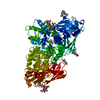
| Title | Crystal Structure of 1,2-beta-oligoglucan phosphorylase from Lachnoclostridium phytofermentans in complex with sophorose, isofagomine, sulfate ion | |||||||||
 Components Components | Uncharacterized protein | |||||||||
 Keywords Keywords | TRANSFERASE / beta-1 / 2-glucan / glycoside phosphorylase | |||||||||
| Function / homology |  Function and homology information Function and homology informationglycosyltransferase activity / carbohydrate metabolic process / metal ion binding Similarity search - Function | |||||||||
| Biological species |  Clostridium phytofermentans ISDg (bacteria) Clostridium phytofermentans ISDg (bacteria) | |||||||||
| Method |  X-RAY DIFFRACTION / SYNCHROTRON / MOLECULAR REPLACEMENT / Resolution: 2 Å X-RAY DIFFRACTION / SYNCHROTRON / MOLECULAR REPLACEMENT / Resolution: 2 Å | |||||||||
 Authors Authors | Nakajima, M. / Tanaka, N. / Furukawa, N. / Nihira, T. / Kodutsumi, Y. / Takahashi, Y. / Sugimoto, N. / Miyanaga, A. / Fushinobu, S. / Taguchi, H. / Nakai, H. | |||||||||
 Citation Citation | Journal: Sci Rep / Year: 2017 Title: Mechanistic insight into the substrate specificity of 1,2-beta-oligoglucan phosphorylase from Lachnoclostridium phytofermentans Authors: Nakajima, M. / Tanaka, N. / Furukawa, N. / Nihira, T. / Kodutsumi, Y. / Takahashi, Y. / Sugimoto, N. / Miyanaga, A. / Fushinobu, S. / Taguchi, H. / Nakai, H. | |||||||||
| History |
|
- Structure visualization
| Structure viewer | Molecule: MolmilJmol/JSmol |
|---|
- Downloads & links
-Download
| PDBx/mmCIF format | 5h41.cif.gz | 466.9 KB | Display | PDBx/mmCIF format |
|---|---|---|---|---|
| PDB format | pdb5h41.ent.gz | 374.2 KB | Display | PDB format |
| PDBx/mmJSON format | 5h41.json.gz | Tree view | PDBx/mmJSON format | |
| Others |  Other downloads Other downloads |
-Validation report
| Arichive directory | https://data.pdbj.org/pub/pdb/validation_reports/h4/5h41ftp://data.pdbj.org/pub/pdb/validation_reports/h4/5h41 | HTTPS FTP |
|---|
-Related structure data
| Related structure data |  5h3zSC  5h40C  5h42C S: Starting model for refinement C: citing same article ( |
|---|---|
| Similar structure data |


-Links
 PDBj
PDBj
- Assembly
| Deposited unit | 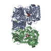
| ||||||||||||||||||
|---|---|---|---|---|---|---|---|---|---|---|---|---|---|---|---|---|---|---|---|
| 1 | 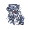
| ||||||||||||||||||
| 2 | 
| ||||||||||||||||||
| Unit cell |
| ||||||||||||||||||
| Noncrystallographic symmetry (NCS) | NCS domain:
NCS domain segments: Component-ID: _ / Ens-ID: 1 / Beg auth comp-ID: GLY / Beg label comp-ID: GLY / End auth comp-ID: ASN / End label comp-ID: ASN / Refine code: _ / Auth seq-ID: 1 - 1113 / Label seq-ID: 2 - 1114
|
-Components
| #1: Protein | Mass: 127590.570 Da / Num. of mol.: 2 Source method: isolated from a genetically manipulated source Source: (gene. exp.) Clostridium phytofermentans ISDg (bacteria)Strain: ISDg / Gene: Cphy_0694 / Production host: #2: Polysaccharide | Source method: isolated from a genetically manipulated source #3: Chemical |   Mass: 147.172 Da / Num. of mol.: 2 / Source method: obtained synthetically / Formula: C6H13NO3 Mass: 147.172 Da / Num. of mol.: 2 / Source method: obtained synthetically / Formula: C6H13NO3#4: Chemical | ChemComp-SO4 /   Mass: 96.063 Da / Num. of mol.: 4 / Source method: obtained synthetically / Formula: SO4 Mass: 96.063 Da / Num. of mol.: 4 / Source method: obtained synthetically / Formula: SO4#5: Water | ChemComp-HOH / |  Mass: 18.015 Da / Num. of mol.: 839 / Source method: isolated from a natural source / Formula: H2O Mass: 18.015 Da / Num. of mol.: 839 / Source method: isolated from a natural source / Formula: H2O |
|---|
-Experimental details
-Experiment
| Experiment | Method: X-RAY DIFFRACTION / Number of used crystals: 1 |
|---|
- Sample preparation
| Crystal | Density Matthews: 2.49 Å3/Da / Density % sol: 50.62 % |
|---|---|
| Crystal grow | Temperature: 298 K / Method: vapor diffusion, hanging drop / pH: 7.5 / Details: PEG3350, calcium acetate, Tris-HCl |
-Data collection
| Diffraction | Mean temperature: 100 K |
|---|---|
| Diffraction source | Source: SYNCHROTRON / Site: Photon Factory  / Beamline: BL-5A / Wavelength: 1 Å / Beamline: BL-5A / Wavelength: 1 Å |
| Detector | Type: ADSC QUANTUM 210r / Detector: CCD / Date: Nov 28, 2015 |
| Radiation | Protocol: SINGLE WAVELENGTH / Monochromatic (M) / Laue (L): M / Scattering type: x-ray |
| Radiation wavelength | Wavelength: 1 Å / Relative weight: 1 |
| Reflection | Resolution: 2→50 Å / Num. obs: 168440 / % possible obs: 96 % / Redundancy: 3.7 % / Rmerge(I) obs: 0.127 / Net I/av σ(I): 1.6 / Net I/σ(I): 3.7 |
| Reflection shell | Resolution: 2→2.03 Å / Redundancy: 3.6 % / Rmerge(I) obs: 0.502 / Mean I/σ(I) obs: 1.6 / Num. measured obs: 29959 / Num. unique all: 8322 / % possible all: 99 |
- Processing
| Software |
| ||||||||||||||||||||||||||||||||||||||||||||||||||||||||||||||||||||||||||||||||||||||||||||||||||||||||||||||||||||||||||||||||||||||||||||||||||||||||||||||||||||||||||||||||||||||
|---|---|---|---|---|---|---|---|---|---|---|---|---|---|---|---|---|---|---|---|---|---|---|---|---|---|---|---|---|---|---|---|---|---|---|---|---|---|---|---|---|---|---|---|---|---|---|---|---|---|---|---|---|---|---|---|---|---|---|---|---|---|---|---|---|---|---|---|---|---|---|---|---|---|---|---|---|---|---|---|---|---|---|---|---|---|---|---|---|---|---|---|---|---|---|---|---|---|---|---|---|---|---|---|---|---|---|---|---|---|---|---|---|---|---|---|---|---|---|---|---|---|---|---|---|---|---|---|---|---|---|---|---|---|---|---|---|---|---|---|---|---|---|---|---|---|---|---|---|---|---|---|---|---|---|---|---|---|---|---|---|---|---|---|---|---|---|---|---|---|---|---|---|---|---|---|---|---|---|---|---|---|---|---|
| Refinement | Method to determine structure: MOLECULAR REPLACEMENT Starting model: 5H3Z Resolution: 2→48.27 Å / Cor.coef. Fo:Fc: 0.96 / Cor.coef. Fo:Fc free: 0.942 / SU B: 4.174 / SU ML: 0.113 / Cross valid method: THROUGHOUT / ESU R: 0.161 / ESU R Free: 0.142 / Stereochemistry target values: MAXIMUM LIKELIHOOD / Details: HYDROGENS HAVE BEEN ADDED IN THE RIDING POSITIONS
| ||||||||||||||||||||||||||||||||||||||||||||||||||||||||||||||||||||||||||||||||||||||||||||||||||||||||||||||||||||||||||||||||||||||||||||||||||||||||||||||||||||||||||||||||||||||
| Solvent computation | Ion probe radii: 0.8 Å / Shrinkage radii: 0.8 Å / VDW probe radii: 1.2 Å / Solvent model: MASK | ||||||||||||||||||||||||||||||||||||||||||||||||||||||||||||||||||||||||||||||||||||||||||||||||||||||||||||||||||||||||||||||||||||||||||||||||||||||||||||||||||||||||||||||||||||||
| Displacement parameters | Biso mean: 27.376 Å2
| ||||||||||||||||||||||||||||||||||||||||||||||||||||||||||||||||||||||||||||||||||||||||||||||||||||||||||||||||||||||||||||||||||||||||||||||||||||||||||||||||||||||||||||||||||||||
| Refinement step | Cycle: 1 / Resolution: 2→48.27 Å
| ||||||||||||||||||||||||||||||||||||||||||||||||||||||||||||||||||||||||||||||||||||||||||||||||||||||||||||||||||||||||||||||||||||||||||||||||||||||||||||||||||||||||||||||||||||||
| Refine LS restraints |
|