 Movie
Movie Controller

[English] 日本語
 Yorodumi
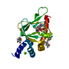
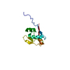
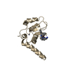
- PDB-5h0o: Crystal structure of deep-sea thermophilic bacteriophage GVE2 HNH... -
+ Open data

- Basic information
| Entry | Database: PDB / ID: 5h0o | ||||||
|---|---|---|---|---|---|---|---|
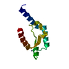
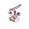
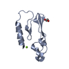
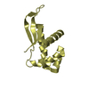
| Title | Crystal structure of deep-sea thermophilic bacteriophage GVE2 HNH endonuclease with manganese ion | ||||||
 Components Components | HNH endonuclease | ||||||
 Keywords Keywords | HYDROLASE / Thermophilic bacteriophage / HNH Endonuclease / DNA nicking | ||||||
| Function / homology |  Function and homology information Function and homology informationendonuclease activity / nucleic acid binding / hydrolase activity / zinc ion binding Similarity search - Function | ||||||
| Biological species |  Geobacillus virus E2 Geobacillus virus E2 | ||||||
| Method |  X-RAY DIFFRACTION / SYNCHROTRON / MOLECULAR REPLACEMENT / Resolution: 1.53 Å X-RAY DIFFRACTION / SYNCHROTRON / MOLECULAR REPLACEMENT / Resolution: 1.53 Å | ||||||
 Authors Authors | Zhang, L.K. / Xu, D.D. / Huang, Y.C. / Gong, Y. | ||||||
 Citation Citation | Journal: Sci Rep / Year: 2017 Title: Structural and functional characterization of deep-sea thermophilic bacteriophage GVE2 HNH endonuclease. Authors: Zhang, L.K. / Xu, D.D. / Huang, Y.C. / Zhu, X.Y. / Rui, M.W. / Wan, T. / Zheng, X. / Shen, Y.L. / Chen, X.D. / Ma, K.S. / Gong, Y. | ||||||
| History |
|
- Structure visualization
| Structure viewer | Molecule: MolmilJmol/JSmol |
|---|
- Downloads & links
-Download
| PDBx/mmCIF format | 5h0o.cif.gz | 62.3 KB | Display | PDBx/mmCIF format |
|---|---|---|---|---|
| PDB format | pdb5h0o.ent.gz | 44.1 KB | Display | PDB format |
| PDBx/mmJSON format | 5h0o.json.gz | Tree view | PDBx/mmJSON format | |
| Others |  Other downloads Other downloads |
-Validation report
| Arichive directory | https://data.pdbj.org/pub/pdb/validation_reports/h0/5h0oftp://data.pdbj.org/pub/pdb/validation_reports/h0/5h0o | HTTPS FTP |
|---|
-Related structure data
| Related structure data |  5h0mSC S: Starting model for refinement C: citing same article ( |
|---|---|
| Similar structure data |
-Links
 PDBj
PDBj
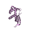
- Assembly
| Deposited unit | 
| ||||||||
|---|---|---|---|---|---|---|---|---|---|
| 1 |
| ||||||||
| Unit cell |
|
-Components
| #1: Protein | Mass: 15754.135 Da / Num. of mol.: 1 Source method: isolated from a genetically manipulated source Source: (gene. exp.) Geobacillus virus E2 / Production host:  |
|---|---|
| #2: Chemical | ChemComp-MN /   Mass: 54.938 Da / Num. of mol.: 1 / Source method: obtained synthetically / Formula: Mn Mass: 54.938 Da / Num. of mol.: 1 / Source method: obtained synthetically / Formula: Mn |
| #3: Water | ChemComp-HOH /  Mass: 18.015 Da / Num. of mol.: 190 / Source method: isolated from a natural source / Formula: H2O Mass: 18.015 Da / Num. of mol.: 190 / Source method: isolated from a natural source / Formula: H2O |
| Sequence details | THE SEQUENCE HAS BEEN DEPOSITED TO DATABASE WITH ACCESSION NUMBER YP_001522898.1. |
-Experimental details
-Experiment
| Experiment | Method: X-RAY DIFFRACTION / Number of used crystals: 1 |
|---|
- Sample preparation
| Crystal | Density Matthews: 1.83 Å3/Da / Density % sol: 32.65 % |
|---|---|
| Crystal grow | Temperature: 293 K / Method: vapor diffusion, sitting drop / pH: 7.5 Details: 0.1M HEPES, 0.2M sodium chloride, pH 7.5, 25% (w/v) polyethylene glycol 4000 |
-Data collection
| Diffraction | Mean temperature: 100 K |
|---|---|
| Diffraction source | Source: SYNCHROTRON / Site: Photon Factory  / Beamline: BL-1A / Wavelength: 1.1 Å / Beamline: BL-1A / Wavelength: 1.1 Å |
| Detector | Type: DECTRIS PILATUS3 2M / Detector: PIXEL / Date: Apr 28, 2016 |
| Radiation | Protocol: SINGLE WAVELENGTH / Monochromatic (M) / Laue (L): M / Scattering type: x-ray |
| Radiation wavelength | Wavelength: 1.1 Å / Relative weight: 1 |
| Reflection | Resolution: 1.53→50 Å / Num. obs: 19106 / % possible obs: 99.8 % / Redundancy: 13.4 % / Biso Wilson estimate: 13.3 Å2 / Rmerge(I) obs: 0.065 / Net I/σ(I): 21.6 |
| Reflection shell | Resolution: 1.53→1.55 Å / Mean I/σ(I) obs: 2.1 / % possible all: 100 |
- Processing
| Software |
| ||||||||||||||||||||||||||||||||||||||||||||||||||||||||
|---|---|---|---|---|---|---|---|---|---|---|---|---|---|---|---|---|---|---|---|---|---|---|---|---|---|---|---|---|---|---|---|---|---|---|---|---|---|---|---|---|---|---|---|---|---|---|---|---|---|---|---|---|---|---|---|---|---|
| Refinement | Method to determine structure: MOLECULAR REPLACEMENT Starting model: 5H0M Resolution: 1.53→25.856 Å / SU ML: 0.13 / Cross valid method: FREE R-VALUE / σ(F): 1.36 / Phase error: 14.36 / Stereochemistry target values: ML
| ||||||||||||||||||||||||||||||||||||||||||||||||||||||||
| Solvent computation | Shrinkage radii: 0.9 Å / VDW probe radii: 1.11 Å / Solvent model: FLAT BULK SOLVENT MODEL | ||||||||||||||||||||||||||||||||||||||||||||||||||||||||
| Refinement step | Cycle: LAST / Resolution: 1.53→25.856 Å
| ||||||||||||||||||||||||||||||||||||||||||||||||||||||||
| Refine LS restraints |
| ||||||||||||||||||||||||||||||||||||||||||||||||||||||||
| LS refinement shell |
|
