 Movie
Movie Controller

[English] 日本語
 Yorodumi
- PDB-5h0i: Structure of OaAEP1 asparaginyl peptide ligase in its proenzyme form -
+ Open data

- Basic information
| Entry | Database: PDB / ID: 5h0i | ||||||
|---|---|---|---|---|---|---|---|
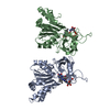
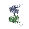
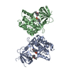
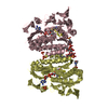
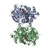
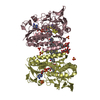
| Title | Structure of OaAEP1 asparaginyl peptide ligase in its proenzyme form | ||||||
 Components Components | Asparaginyl endopeptidase | ||||||
 Keywords Keywords | HYDROLASE | ||||||
| Function / homology |  Function and homology information Function and homology informationlegumain / vacuolar protein processing / vacuole / proteolysis involved in protein catabolic process / cysteine-type endopeptidase activity Similarity search - Function | ||||||
| Biological species |  Oldenlandia affinis (plant) Oldenlandia affinis (plant) | ||||||
| Method |  X-RAY DIFFRACTION / SYNCHROTRON / MOLECULAR REPLACEMENT / Resolution: 2.56 Å X-RAY DIFFRACTION / SYNCHROTRON / MOLECULAR REPLACEMENT / Resolution: 2.56 Å | ||||||
 Authors Authors | Yang, R. / Wong, Y.H. / Lescar, J. / Wu, B. | ||||||
| Funding support |  Singapore, 1items Singapore, 1items
| ||||||
 Citation Citation | Journal: J. Am. Chem. Soc. / Year: 2017 Title: Engineering a Catalytically Efficient Recombinant Protein Ligase Authors: Yang, R. / Wong, Y.H. / Nguyen, G.K.T. / Tam, J.P. / Lescar, J. / Wu, B. | ||||||
| History |
|
- Structure visualization
| Structure viewer | Molecule: MolmilJmol/JSmol |
|---|
- Downloads & links
-Download
| PDBx/mmCIF format | 5h0i.cif.gz | 330.7 KB | Display | PDBx/mmCIF format |
|---|---|---|---|---|
| PDB format | pdb5h0i.ent.gz | 268.6 KB | Display | PDB format |
| PDBx/mmJSON format | 5h0i.json.gz | Tree view | PDBx/mmJSON format | |
| Others |  Other downloads Other downloads |
-Validation report
| Summary document | 5h0i_validation.pdf.gz | 435.3 KB | Display | wwPDB validaton report |
|---|---|---|---|---|
| Full document | 5h0i_full_validation.pdf.gz | 445.6 KB | Display | |
| Data in XML | 5h0i_validation.xml.gz | 31.7 KB | Display | |
| Data in CIF | 5h0i_validation.cif.gz | 45.4 KB | Display | |
| Arichive directory | https://data.pdbj.org/pub/pdb/validation_reports/h0/5h0iftp://data.pdbj.org/pub/pdb/validation_reports/h0/5h0i | HTTPS FTP |
-Related structure data
| Related structure data |  4nokS S: Starting model for refinement |
|---|---|
| Similar structure data |


-Links
 PDBj
PDBj
- Assembly
| Deposited unit | 
| ||||||||
|---|---|---|---|---|---|---|---|---|---|
| 1 |
| ||||||||
| Unit cell |
|
-Components
| #1: Protein | Mass: 49393.871 Da / Num. of mol.: 2 / Fragment: UNP residues 24-474 Source method: isolated from a genetically manipulated source Source: (gene. exp.) Oldenlandia affinis (plant) / Production host:  #2: Water | ChemComp-HOH / |  Mass: 18.015 Da / Num. of mol.: 292 / Source method: isolated from a natural source / Formula: H2O Mass: 18.015 Da / Num. of mol.: 292 / Source method: isolated from a natural source / Formula: H2OHas protein modification | Y | |
|---|
-Experimental details
-Experiment
| Experiment | Method: X-RAY DIFFRACTION / Number of used crystals: 1 |
|---|
- Sample preparation
| Crystal | Density Matthews: 2.73 Å3/Da / Density % sol: 54.87 % |
|---|---|
| Crystal grow | Temperature: 290 K / Method: vapor diffusion, hanging drop / pH: 4.5 Details: 200mM NH4NO3, pH ~ 4.5, 13-15%(w/v) PEG 3,350 with 10%(v/v) glycerol or ethylene glycol |
-Data collection
| Diffraction | Mean temperature: 77 K |
|---|---|
| Diffraction source | Source: SYNCHROTRON / Site: SLS  / Beamline: X06SA / Wavelength: 1 Å / Beamline: X06SA / Wavelength: 1 Å |
| Detector | Type: DECTRIS PILATUS 6M / Detector: PIXEL / Date: Jun 17, 2016 |
| Radiation | Protocol: SINGLE WAVELENGTH / Monochromatic (M) / Laue (L): M / Scattering type: x-ray |
| Radiation wavelength | Wavelength: 1 Å / Relative weight: 1 |
| Reflection | Resolution: 2.56→71.72 Å / Num. obs: 34420 / % possible obs: 99.7 % / Redundancy: 3 % / Biso Wilson estimate: 63.02 Å2 / Rmerge(I) obs: 0.095 / Net I/σ(I): 7 |
| Reflection shell | Resolution: 2.56→2.7 Å / Redundancy: 2.8 % / Rmerge(I) obs: 0.7 / Mean I/σ(I) obs: 1.7 / % possible all: 99.6 |
- Processing
| Software |
| ||||||||||||||||||||||||||||||||||||||||||||||||||||||||||||||||||||||||||||||||||||||||||||||||||||||||||||||||||
|---|---|---|---|---|---|---|---|---|---|---|---|---|---|---|---|---|---|---|---|---|---|---|---|---|---|---|---|---|---|---|---|---|---|---|---|---|---|---|---|---|---|---|---|---|---|---|---|---|---|---|---|---|---|---|---|---|---|---|---|---|---|---|---|---|---|---|---|---|---|---|---|---|---|---|---|---|---|---|---|---|---|---|---|---|---|---|---|---|---|---|---|---|---|---|---|---|---|---|---|---|---|---|---|---|---|---|---|---|---|---|---|---|---|---|---|
| Refinement | Method to determine structure: MOLECULAR REPLACEMENT Starting model: 4NOK Resolution: 2.56→71.72 Å / Cor.coef. Fo:Fc: 0.934 / Cor.coef. Fo:Fc free: 0.901 / Rfactor Rfree error: 0.01 / SU R Cruickshank DPI: 0.419 / Cross valid method: THROUGHOUT / σ(F): 0 / SU R Blow DPI: 0.427 / SU Rfree Blow DPI: 0.247 / SU Rfree Cruickshank DPI: 0.249
| ||||||||||||||||||||||||||||||||||||||||||||||||||||||||||||||||||||||||||||||||||||||||||||||||||||||||||||||||||
| Displacement parameters | Biso mean: 68.81 Å2
| ||||||||||||||||||||||||||||||||||||||||||||||||||||||||||||||||||||||||||||||||||||||||||||||||||||||||||||||||||
| Refine analyze | Luzzati coordinate error obs: 0.31 Å | ||||||||||||||||||||||||||||||||||||||||||||||||||||||||||||||||||||||||||||||||||||||||||||||||||||||||||||||||||
| Refinement step | Cycle: 1 / Resolution: 2.56→71.72 Å
| ||||||||||||||||||||||||||||||||||||||||||||||||||||||||||||||||||||||||||||||||||||||||||||||||||||||||||||||||||
| Refine LS restraints |
| ||||||||||||||||||||||||||||||||||||||||||||||||||||||||||||||||||||||||||||||||||||||||||||||||||||||||||||||||||
| LS refinement shell | Resolution: 2.56→2.64 Å / Rfactor Rfree error: 0 / Total num. of bins used: 17
| ||||||||||||||||||||||||||||||||||||||||||||||||||||||||||||||||||||||||||||||||||||||||||||||||||||||||||||||||||
| Refinement TLS params. | Method: refined / Refine-ID: X-RAY DIFFRACTION
| ||||||||||||||||||||||||||||||||||||||||||||||||||||||||||||||||||||||||||||||||||||||||||||||||||||||||||||||||||
| Refinement TLS group |
|
