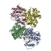
 Movie
Movie Controller


+ Open data

- Basic information
| Entry | Database: PDB / ID: 5gzs | ||||||
|---|---|---|---|---|---|---|---|
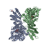
| Title | Structure of VC protein | ||||||
 Components Components | GGDEF family protein | ||||||
 Keywords Keywords | SIGNALING PROTEIN / the periplasmic portion / CdgH / Vibrio cholerae / diguanylate cyclase | ||||||
| Function / homology |  Function and homology information Function and homology information | ||||||
| Biological species |   Vibrio cholerae (bacteria) Vibrio cholerae (bacteria) | ||||||
| Method |  X-RAY DIFFRACTION / SYNCHROTRON / Resolution: 2.601 Å X-RAY DIFFRACTION / SYNCHROTRON / Resolution: 2.601 Å | ||||||
 Authors Authors | Xu, M. / Wang, Y.Z. / Yang, X.A. / Xie, W. / Jiang, T. | ||||||
 Citation Citation | Journal: Sci Rep / Year: 2017 Title: Structural studies of the periplasmic portion of the diguanylate cyclase CdgH from Vibrio cholerae. Authors: Xu, M. / Wang, Y.Z. / Yang, X.A. / Jiang, T. / Xie, W. | ||||||
| History |
|
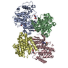
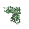
- Structure visualization
| Structure viewer | Molecule: MolmilJmol/JSmol |
|---|
- Downloads & links
-Download
| PDBx/mmCIF format | 5gzs.cif.gz | 191.9 KB | Display | PDBx/mmCIF format |
|---|---|---|---|---|
| PDB format | pdb5gzs.ent.gz | 152.9 KB | Display | PDB format |
| PDBx/mmJSON format | 5gzs.json.gz | Tree view | PDBx/mmJSON format | |
| Others |  Other downloads Other downloads |
-Validation report
| Arichive directory | https://data.pdbj.org/pub/pdb/validation_reports/gz/5gzsftp://data.pdbj.org/pub/pdb/validation_reports/gz/5gzs | HTTPS FTP |
|---|
-Related structure data
| Similar structure data |
|---|


-Links
 PDBj
PDBj


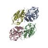
- Assembly
| Deposited unit | 
| |||||||||
|---|---|---|---|---|---|---|---|---|---|---|
| 1 | 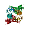
| |||||||||
| Unit cell |
| |||||||||
| Components on special symmetry positions |
|
-Components
| #1: Protein | Mass: 51217.062 Da / Num. of mol.: 1 / Fragment: UNP RESIDUES 26-471 Source method: isolated from a genetically manipulated source Source: (gene. exp.) Vibrio cholerae (bacteria) / Gene: ERS013200_00600 / Production host: |
|---|---|
| #2: Chemical | ChemComp-ARG /   Type: L-peptide linking / Mass: 175.209 Da / Num. of mol.: 1 Type: L-peptide linking / Mass: 175.209 Da / Num. of mol.: 1Source method: isolated from a genetically manipulated source Formula: C6H15N4O2 |
| #3: Water | ChemComp-HOH /  Mass: 18.015 Da / Num. of mol.: 74 / Source method: isolated from a natural source / Formula: H2O Mass: 18.015 Da / Num. of mol.: 74 / Source method: isolated from a natural source / Formula: H2O |
| Has protein modification | Y |
-Experimental details
-Experiment
| Experiment | Method: X-RAY DIFFRACTION / Number of used crystals: 1 |
|---|
- Sample preparation
| Crystal | Density Matthews: 2.44 Å3/Da / Density % sol: 49.59 % |
|---|---|
| Crystal grow | Temperature: 293 K / Method: vapor diffusion, hanging drop / pH: 8 Details: 0.1 M Tris-HCl (pH 8.0), 10% w/v polyethylene glycol 8000 |
-Data collection
| Diffraction | Mean temperature: 100 K |
|---|---|
| Diffraction source | Source: SYNCHROTRON / Site: SSRF  / Beamline: BL18U1 / Wavelength: 0.97848 Å / Beamline: BL18U1 / Wavelength: 0.97848 Å |
| Detector | Type: DECTRIS PILATUS3 6M / Detector: PIXEL / Date: May 1, 2015 |
| Radiation | Protocol: SINGLE WAVELENGTH / Monochromatic (M) / Laue (L): M / Scattering type: x-ray |
| Radiation wavelength | Wavelength: 0.97848 Å / Relative weight: 1 |
| Reflection | Resolution: 2.601→40 Å / Num. obs: 16125 / % possible obs: 99 % / Redundancy: 10.5 % / Net I/σ(I): 28.9 |
- Processing
| Software |
| |||||||||||||||||||||||||||||||||||||||||||||||||
|---|---|---|---|---|---|---|---|---|---|---|---|---|---|---|---|---|---|---|---|---|---|---|---|---|---|---|---|---|---|---|---|---|---|---|---|---|---|---|---|---|---|---|---|---|---|---|---|---|---|---|
| Refinement | Resolution: 2.601→40 Å / SU ML: 0.33 / Cross valid method: FREE R-VALUE / σ(F): 1.34 / Phase error: 29.07
| |||||||||||||||||||||||||||||||||||||||||||||||||
| Solvent computation | Shrinkage radii: 0.9 Å / VDW probe radii: 1.11 Å | |||||||||||||||||||||||||||||||||||||||||||||||||
| Refinement step | Cycle: LAST / Resolution: 2.601→40 Å
| |||||||||||||||||||||||||||||||||||||||||||||||||
| Refine LS restraints |
| |||||||||||||||||||||||||||||||||||||||||||||||||
| LS refinement shell |
|
