| Entry | Database: PDB / ID: 5gpj
|
|---|
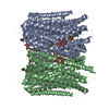
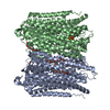
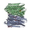
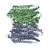
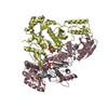
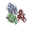
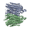
| Title | Crystal Structure of Proton-Pumping Pyrophosphatase |
|---|
 Components Components | Pyrophosphate-energized vacuolar membrane proton pump |
|---|
 Keywords Keywords | HYDROLASE / Vigna radiata / Proton-Pumping / Phosphate-bound |
|---|
| Function / homology | H+-exporting diphosphatase / diphosphate hydrolysis-driven proton transmembrane transporter activity / Pyrophosphate-energised proton pump / Inorganic H+ pyrophosphatase / inorganic diphosphate phosphatase activity / vacuolar membrane / metal ion binding / PHOSPHATE ION / Pyrophosphate-energized vacuolar membrane proton pump Function and homology information Function and homology information |
|---|
| Biological species |  Vigna radiata var. radiata (mung bean) Vigna radiata var. radiata (mung bean) |
|---|
| Method |  X-RAY DIFFRACTION / SYNCHROTRON / MOLECULAR REPLACEMENT / Resolution: 3.5 Å X-RAY DIFFRACTION / SYNCHROTRON / MOLECULAR REPLACEMENT / Resolution: 3.5 Å |
|---|
 Authors Authors | Li, K.M. / Tsai, J.Y. / Sun, Y.J. |
|---|
| Funding support |  Taiwan, 2items Taiwan, 2items | Organization | Grant number | Country |
|---|
| Ministry of Science and Technolgy | 101-2311-B-007-009-MY3 and 103-2627-M-007-008 | Taiwan | | Academia Sinica | AS-103-TP-B11 | Taiwan |
|
|---|
 Citation Citation | Journal: Nat Commun / Year: 2016
Title: Membrane pyrophosphatases from Thermotoga maritima and Vigna radiata suggest a conserved coupling mechanism
Authors: Li, K.M. / Wilkinson, C. / Kellosalo, J. / Tsai, J.Y. / Kajander, T. / Jeuken, L.J. / Sun, Y.J. / Goldman, A. |
|---|
| History | | Deposition | Aug 3, 2016 | Deposition site: PDBJ / Processing site: PDBJ |
|---|
| Revision 1.0 | Dec 28, 2016 | Provider: repository / Type: Initial release |
|---|
| Revision 1.1 | Nov 8, 2023 | Group: Data collection / Database references ...Data collection / Database references / Derived calculations / Refinement description
Category: chem_comp_atom / chem_comp_bond ...chem_comp_atom / chem_comp_bond / database_2 / pdbx_initial_refinement_model / struct_conn
Item: _database_2.pdbx_DOI / _database_2.pdbx_database_accession ..._database_2.pdbx_DOI / _database_2.pdbx_database_accession / _struct_conn.pdbx_dist_value / _struct_conn.ptnr1_auth_asym_id / _struct_conn.ptnr1_auth_comp_id / _struct_conn.ptnr1_auth_seq_id / _struct_conn.ptnr1_label_asym_id / _struct_conn.ptnr1_label_atom_id / _struct_conn.ptnr1_label_comp_id / _struct_conn.ptnr1_label_seq_id / _struct_conn.ptnr2_auth_asym_id / _struct_conn.ptnr2_auth_seq_id / _struct_conn.ptnr2_label_asym_id |
|---|
| Revision 1.2 | Oct 16, 2024 | Group: Structure summary / Category: pdbx_entry_details / pdbx_modification_feature |
|---|
|
|---|
 Movie
Movie Controller


 Open data
 Basic information
 Structure visualization
 Downloads & links
 Other downloads
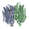


 PDBj
PDBj Assembly


 Mass: 94.971 Da / Num. of mol.: 4 / Source method: obtained synthetically / Formula: PO4
 Mass: 24.305 Da / Num. of mol.: 8 / Source method: obtained synthetically / Formula: Mg
 Sample preparation
 Processing