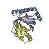
Entry Database : PDB / ID : 5gnvTitle Structure of PSD-95/MAP1A complex reveals unique target recognition mode of MAGUK GK domain Disks large homolog 4 Microtubule-associated protein 1A Keywords / Function / homology Function Domain/homology Component
/ / / / / / / / / / / / / / / / / / / / / / / / / / / / / / / / / / / / / / / / / / / / / / / / / / / / / / / / / / / / / / / / / / / / / / / / / / / / / / / / / / / / / / / / / / / / / / / / / / / / / / / / / / / / / / / / / / / / / / / / / / / / / / / / / / / / / / / / / / / / / / / / / / / Biological species Rattus norvegicus (Norway rat)Mus musculus (house mouse)Method / / / Resolution : 2.596 Å Authors Shang, Y. / Xia, Y. / Zhu, R. / Zhu, J. Funding support Organization Grant number Country National Natural Science Foundation of China 31470733 the Shanghai YangFan Plan for Young Scientists 14YF1406700 the SIBS Frontier Science Program for talented young scientists 2014KIP101
Journal : Biochem. J. / Year : 2017Title : Structure of the PSD-95/MAP1A complex reveals a unique target recognition mode of the MAGUK GK domainAuthors : Xia, Y. / Shang, Y. / Zhang, R. / Zhu, J. History Deposition Jul 25, 2016 Deposition site / Processing site Revision 1.0 Aug 2, 2017 Provider / Type Revision 1.1 Nov 1, 2017 Group / Category / citation_authorItem _citation.country / _citation.journal_abbrev ... _citation.country / _citation.journal_abbrev / _citation.journal_id_ASTM / _citation.journal_id_CSD / _citation.journal_id_ISSN / _citation.journal_volume / _citation.page_first / _citation.page_last / _citation.pdbx_database_id_DOI / _citation.pdbx_database_id_PubMed / _citation.title / _citation.year / _citation_author.name Revision 1.2 Mar 20, 2024 Group / Database references / Derived calculationsCategory chem_comp_atom / chem_comp_bond ... chem_comp_atom / chem_comp_bond / database_2 / pdbx_struct_special_symmetry Item / _database_2.pdbx_database_accession
Show all Show less
 Movie
Movie Controller

 Yorodumi
 Open data
 Basic information
 Components
 Keywords
 Function and homology information
 X-RAY DIFFRACTION /
  Authors
 China, 3items
  Citation
 Structure visualization
 Downloads & links
 Other downloads
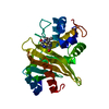


 PDBj
PDBj


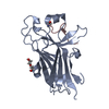
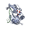
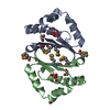
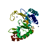
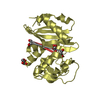
 Assembly
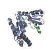


 Mass: 96.063 Da / Num. of mol.: 2 / Source method: obtained synthetically / Formula: SO4
 Mass: 18.015 Da / Num. of mol.: 36 / Source method: isolated from a natural source / Formula: H2O
 Sample preparation
 Processing