 Movie
Movie Controller


+ Open data

- Basic information
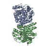
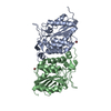
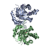
| Entry | Database: PDB / ID: 5gl4 | ||||||
|---|---|---|---|---|---|---|---|
| Title | Crystal structure of TON_0340 in complex with Mn | ||||||
 Components Components | Uncharacterized protein | ||||||
 Keywords Keywords | UNKNOWN FUNCTION / TON_0340 / Thermococcus onnurineus / Mn2+-dependent phosphatase | ||||||
| Function / homology |  Function and homology information Function and homology informationD-glutamate cyclase activity / glutamate metabolic process / metal ion binding Similarity search - Function | ||||||
| Biological species |   Thermococcus onnurineus (archaea) Thermococcus onnurineus (archaea) | ||||||
| Method |  X-RAY DIFFRACTION / SYNCHROTRON / MOLECULAR REPLACEMENT / Resolution: 2.2 Å X-RAY DIFFRACTION / SYNCHROTRON / MOLECULAR REPLACEMENT / Resolution: 2.2 Å | ||||||
 Authors Authors | Lee, S.G. / Sohn, Y.S. / Oh, B.H. | ||||||
| Funding support |  Korea, Republic Of, 1items Korea, Republic Of, 1items
| ||||||
 Citation Citation | Journal: PLoS ONE / Year: 2016 Title: Identification of a Highly Conserved Hypothetical Protein TON_0340 as a Probable Manganese-Dependent Phosphatase. Authors: Sohn, Y.S. / Lee, S.G. / Lee, K.H. / Ku, B. / Shin, H.C. / Cha, S.S. / Kim, Y.G. / Lee, H.S. / Kang, S.G. / Oh, B.H. #1: Journal: Acta Crystallogr. D Biol. Crystallogr. / Year: 2012Title: Experimental phasing using zinc anomalous scattering. Authors: Cha, S.S. / An, Y.J. / Jeong, C.S. / Kim, M.K. / Lee, S.G. / Lee, K.H. / Oh, B.H. | ||||||
| History |
|
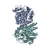
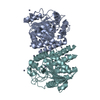
- Structure visualization
| Structure viewer | Molecule: MolmilJmol/JSmol |
|---|
- Downloads & links
-Download
| PDBx/mmCIF format | 5gl4.cif.gz | 307.3 KB | Display | PDBx/mmCIF format |
|---|---|---|---|---|
| PDB format | pdb5gl4.ent.gz | 250.9 KB | Display | PDB format |
| PDBx/mmJSON format | 5gl4.json.gz | Tree view | PDBx/mmJSON format | |
| Others |  Other downloads Other downloads |
-Validation report
| Arichive directory | https://data.pdbj.org/pub/pdb/validation_reports/gl/5gl4ftp://data.pdbj.org/pub/pdb/validation_reports/gl/5gl4 | HTTPS FTP |
|---|
-Related structure data
| Related structure data | 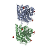 5gkxC 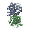 5gl2C 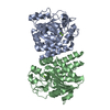 5gl3C  4fc5S S: Starting model for refinement C: citing same article ( |
|---|---|
| Similar structure data |


-Links
 PDBj
PDBj- Assembly
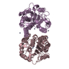
| Deposited unit | 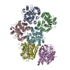
| ||||||||
|---|---|---|---|---|---|---|---|---|---|
| 1 | 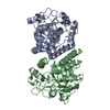
| ||||||||
| 2 | 
| ||||||||
| 3 | 
| ||||||||
| Unit cell |
|
-Components
| #1: Protein | Mass: 29107.531 Da / Num. of mol.: 6 Source method: isolated from a genetically manipulated source Source: (gene. exp.) Thermococcus onnurineus (strain NA1) (archaea)Strain: NA1 / Gene: TON_0340 / Production host:  #2: Chemical | ChemComp-MN /   Mass: 54.938 Da / Num. of mol.: 17 / Source method: obtained synthetically / Formula: Mn Mass: 54.938 Da / Num. of mol.: 17 / Source method: obtained synthetically / Formula: Mn#3: Water | ChemComp-HOH / |  Mass: 18.015 Da / Num. of mol.: 262 / Source method: isolated from a natural source / Formula: H2O Mass: 18.015 Da / Num. of mol.: 262 / Source method: isolated from a natural source / Formula: H2O |
|---|
-Experimental details
-Experiment
| Experiment | Method: X-RAY DIFFRACTION / Number of used crystals: 1 |
|---|
- Sample preparation
| Crystal | Density Matthews: 2.97 Å3/Da / Density % sol: 58.64 % |
|---|---|
| Crystal grow | Temperature: 295 K / Method: vapor diffusion, hanging drop Details: sodium acetate pH, 16% 2-methyl-2,4-pentanediol, manganese chloride |
-Data collection
| Diffraction | Mean temperature: 295 K | ||||||||||||||||||||||||||||||||||||||||||||||||||||||||||||||||||||||||||||||||||||||||||||||||||||||||||||||||||||||||||||||
|---|---|---|---|---|---|---|---|---|---|---|---|---|---|---|---|---|---|---|---|---|---|---|---|---|---|---|---|---|---|---|---|---|---|---|---|---|---|---|---|---|---|---|---|---|---|---|---|---|---|---|---|---|---|---|---|---|---|---|---|---|---|---|---|---|---|---|---|---|---|---|---|---|---|---|---|---|---|---|---|---|---|---|---|---|---|---|---|---|---|---|---|---|---|---|---|---|---|---|---|---|---|---|---|---|---|---|---|---|---|---|---|---|---|---|---|---|---|---|---|---|---|---|---|---|---|---|---|
| Diffraction source | Source: SYNCHROTRON / Site: Photon Factory  / Beamline: AR-NW12A / Wavelength: 1 Å / Beamline: AR-NW12A / Wavelength: 1 Å | ||||||||||||||||||||||||||||||||||||||||||||||||||||||||||||||||||||||||||||||||||||||||||||||||||||||||||||||||||||||||||||||
| Detector | Type: ADSC QUANTUM 210 / Detector: CCD / Date: Dec 5, 2011 | ||||||||||||||||||||||||||||||||||||||||||||||||||||||||||||||||||||||||||||||||||||||||||||||||||||||||||||||||||||||||||||||
| Radiation | Protocol: SINGLE WAVELENGTH / Monochromatic (M) / Laue (L): M / Scattering type: x-ray | ||||||||||||||||||||||||||||||||||||||||||||||||||||||||||||||||||||||||||||||||||||||||||||||||||||||||||||||||||||||||||||||
| Radiation wavelength | Wavelength: 1 Å / Relative weight: 1 | ||||||||||||||||||||||||||||||||||||||||||||||||||||||||||||||||||||||||||||||||||||||||||||||||||||||||||||||||||||||||||||||
| Reflection | Resolution: 2.03→50 Å / Num. obs: 112616 / % possible obs: 82.3 % / Redundancy: 3.9 % / Rmerge(I) obs: 0.063 / Χ2: 2.004 / Net I/av σ(I): 19.88 / Net I/σ(I): 15 / Num. measured all: 443354 | ||||||||||||||||||||||||||||||||||||||||||||||||||||||||||||||||||||||||||||||||||||||||||||||||||||||||||||||||||||||||||||||
| Reflection shell | Diffraction-ID: 1 / Rejects: _
|
- Processing
| Software |
| ||||||||||||||||||||||||
|---|---|---|---|---|---|---|---|---|---|---|---|---|---|---|---|---|---|---|---|---|---|---|---|---|---|
| Refinement | Method to determine structure: MOLECULAR REPLACEMENT Starting model: 4FC5 Resolution: 2.2→50 Å / Cross valid method: FREE R-VALUE / σ(F): 38
| ||||||||||||||||||||||||
| Solvent computation | Bsol: 27.5887 Å2 | ||||||||||||||||||||||||
| Displacement parameters | Biso max: 121.85 Å2 / Biso mean: 41.3897 Å2 / Biso min: 15.42 Å2
| ||||||||||||||||||||||||
| Refinement step | Cycle: final / Resolution: 2.2→50 Å
| ||||||||||||||||||||||||
| Refine LS restraints |
| ||||||||||||||||||||||||
| Xplor file |
|
