 Movie
Movie Controller


+ Open data

- Basic information
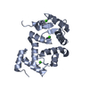
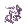
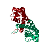
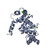
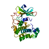
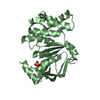
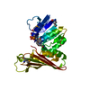
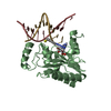
| Entry | Database: PDB / ID: 5g4p | ||||||
|---|---|---|---|---|---|---|---|
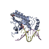
| Title | Crystal structure of human hippocalcin at 2.4 A resolution | ||||||
 Components Components | Neuron-specific calcium-binding protein hippocalcin | ||||||
 Keywords Keywords | CALCIUM BINDING PROTEIN / CA BINDING PROTEIN / CALCIUM-BINDING PROTEIN | ||||||
| Function / homology |  Function and homology information Function and homology informationregulation of voltage-gated calcium channel activity / response to Aroclor 1254 / response to ketamine / cellular response to electrical stimulus / cellular response to L-glutamate / dendritic spine head / regulation of postsynaptic neurotransmitter receptor internalization / neuronal cell body membrane / inner ear development / regulation of signal transduction ...regulation of voltage-gated calcium channel activity / response to Aroclor 1254 / response to ketamine / cellular response to electrical stimulus / cellular response to L-glutamate / dendritic spine head / regulation of postsynaptic neurotransmitter receptor internalization / neuronal cell body membrane / inner ear development / regulation of signal transduction / positive regulation of protein targeting to membrane / cellular response to calcium ion / dendrite membrane / dendrite cytoplasm / calcium-mediated signaling / kinase binding / actin binding / retina development in camera-type eye / perikaryon / axon / calcium ion binding / glutamatergic synapse / membrane / identical protein binding / cytoplasm / cytosol Similarity search - Function | ||||||
| Biological species |  Homo sapiens (human) Homo sapiens (human) | ||||||
| Method |  X-RAY DIFFRACTION / SYNCHROTRON / MOLECULAR REPLACEMENT / Resolution: 2.42 Å X-RAY DIFFRACTION / SYNCHROTRON / MOLECULAR REPLACEMENT / Resolution: 2.42 Å | ||||||
 Authors Authors | Antonyuk, S.V. / Helassa, N. / Lian, L.Y. / Haynes, L.P. / Burgoyne, R.D. | ||||||
 Citation Citation | Journal: Hum. Mol. Genet. / Year: 2017 Title: Biophysical and functional characterization of hippocalcin mutants responsible for human dystonia. Authors: Helassa, N. / Antonyuk, S.V. / Lian, L.Y. / Haynes, L.P. / Burgoyne, R.D. | ||||||
| History |
|
- Structure visualization
| Structure viewer | Molecule: MolmilJmol/JSmol |
|---|
- Downloads & links
-Download
| PDBx/mmCIF format | 5g4p.cif.gz | 163.6 KB | Display | PDBx/mmCIF format |
|---|---|---|---|---|
| PDB format | pdb5g4p.ent.gz | 131.8 KB | Display | PDB format |
| PDBx/mmJSON format | 5g4p.json.gz | Tree view | PDBx/mmJSON format | |
| Others |  Other downloads Other downloads |
-Validation report
| Arichive directory | https://data.pdbj.org/pub/pdb/validation_reports/g4/5g4pftp://data.pdbj.org/pub/pdb/validation_reports/g4/5g4p | HTTPS FTP |
|---|
-Related structure data
| Related structure data |  5g58C  5m6cC  5aeqS S: Starting model for refinement C: citing same article ( |
|---|---|
| Similar structure data |
-Links
 PDBj
PDBj

- Assembly
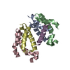
| Deposited unit | 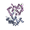
| |||||||||||||||||||||||||||||||||||||||
|---|---|---|---|---|---|---|---|---|---|---|---|---|---|---|---|---|---|---|---|---|---|---|---|---|---|---|---|---|---|---|---|---|---|---|---|---|---|---|---|---|
| 1 | 
| |||||||||||||||||||||||||||||||||||||||
| 2 | 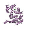
| |||||||||||||||||||||||||||||||||||||||
| Unit cell |
| |||||||||||||||||||||||||||||||||||||||
| Noncrystallographic symmetry (NCS) | NCS domain:
NCS domain segments:
NCS oper:
|
-Components
| #1: Protein | Mass: 22453.402 Da / Num. of mol.: 2 / Mutation: YES Source method: isolated from a genetically manipulated source Source: (gene. exp.) Homo sapiens (human) / Gene: HPCA, BDR2 / Production host:  #2: Chemical | ChemComp-CA /   Mass: 40.078 Da / Num. of mol.: 6 / Source method: obtained synthetically / Formula: Ca Mass: 40.078 Da / Num. of mol.: 6 / Source method: obtained synthetically / Formula: Ca#3: Water | ChemComp-HOH / |  Mass: 18.015 Da / Num. of mol.: 34 / Source method: isolated from a natural source / Formula: H2O Mass: 18.015 Da / Num. of mol.: 34 / Source method: isolated from a natural source / Formula: H2O |
|---|
-Experimental details
-Experiment
| Experiment | Method: X-RAY DIFFRACTION / Number of used crystals: 1 |
|---|
- Sample preparation
| Crystal | Density Matthews: 2.38 Å3/Da / Density % sol: 48 % / Description: NONE |
|---|---|
| Crystal grow | pH: 5.5 Details: 0.1 M SODIUM CITRATE TRIBASIC DIHYDRATE PH 5.5, 18% V/V 2-PROPANOL, 16% W/V PEG 4000 |
-Data collection
| Diffraction | Mean temperature: 100 K |
|---|---|
| Diffraction source | Source: SYNCHROTRON / Site: Diamond  / Beamline: I02 / Wavelength: 0.98 / Beamline: I02 / Wavelength: 0.98 |
| Detector | Type: DECTRIS PILATUS 6M / Detector: PIXEL / Date: Jan 21, 2016 / Details: MIRRORS |
| Radiation | Monochromator: SI111 / Protocol: SINGLE WAVELENGTH / Monochromatic (M) / Laue (L): M / Scattering type: x-ray |
| Radiation wavelength | Wavelength: 0.98 Å / Relative weight: 1 |
| Reflection | Resolution: 2.42→43.56 Å / Num. obs: 15734 / % possible obs: 98.8 % / Redundancy: 3.3 % / Biso Wilson estimate: 63.7 Å2 / Rmerge(I) obs: 0.04 / Net I/σ(I): 11.07 |
| Reflection shell | Resolution: 2.42→2.59 Å / Redundancy: 3.2 % / Rmerge(I) obs: 0.62 / Mean I/σ(I) obs: 1.8 / % possible all: 99.9 |
- Processing
| Software |
| ||||||||||||||||||||||||||||||||||||||||||||||||||||||||||||||||||||||||||||||||||||||||||||||||||||||||||||||||||||||||||||||||||||||||||||||||||||||||||||||||||||||||||||||||||||||
|---|---|---|---|---|---|---|---|---|---|---|---|---|---|---|---|---|---|---|---|---|---|---|---|---|---|---|---|---|---|---|---|---|---|---|---|---|---|---|---|---|---|---|---|---|---|---|---|---|---|---|---|---|---|---|---|---|---|---|---|---|---|---|---|---|---|---|---|---|---|---|---|---|---|---|---|---|---|---|---|---|---|---|---|---|---|---|---|---|---|---|---|---|---|---|---|---|---|---|---|---|---|---|---|---|---|---|---|---|---|---|---|---|---|---|---|---|---|---|---|---|---|---|---|---|---|---|---|---|---|---|---|---|---|---|---|---|---|---|---|---|---|---|---|---|---|---|---|---|---|---|---|---|---|---|---|---|---|---|---|---|---|---|---|---|---|---|---|---|---|---|---|---|---|---|---|---|---|---|---|---|---|---|---|
| Refinement | Method to determine structure: MOLECULAR REPLACEMENT Starting model: PDB ENTRY 5AEQ Resolution: 2.42→47.43 Å / Cor.coef. Fo:Fc: 0.958 / Cor.coef. Fo:Fc free: 0.944 / SU B: 36.292 / SU ML: 0.37 / Cross valid method: THROUGHOUT / ESU R: 0.573 / ESU R Free: 0.308 / Stereochemistry target values: MAXIMUM LIKELIHOOD / Details: HYDROGENS HAVE BEEN ADDED IN THE RIDING POSITIONS
| ||||||||||||||||||||||||||||||||||||||||||||||||||||||||||||||||||||||||||||||||||||||||||||||||||||||||||||||||||||||||||||||||||||||||||||||||||||||||||||||||||||||||||||||||||||||
| Solvent computation | Ion probe radii: 0.8 Å / Shrinkage radii: 0.8 Å / VDW probe radii: 1.2 Å / Solvent model: MASK | ||||||||||||||||||||||||||||||||||||||||||||||||||||||||||||||||||||||||||||||||||||||||||||||||||||||||||||||||||||||||||||||||||||||||||||||||||||||||||||||||||||||||||||||||||||||
| Displacement parameters | Biso mean: 78.609 Å2
| ||||||||||||||||||||||||||||||||||||||||||||||||||||||||||||||||||||||||||||||||||||||||||||||||||||||||||||||||||||||||||||||||||||||||||||||||||||||||||||||||||||||||||||||||||||||
| Refinement step | Cycle: 1 / Resolution: 2.42→47.43 Å
| ||||||||||||||||||||||||||||||||||||||||||||||||||||||||||||||||||||||||||||||||||||||||||||||||||||||||||||||||||||||||||||||||||||||||||||||||||||||||||||||||||||||||||||||||||||||
| Refine LS restraints |
|