 Movie
Movie Controller

[English] 日本語
 Yorodumi
- PDB-5ft5: Crystal structure of the cysteine desulfurase CsdA (persulfurated... -
+ Open data

- Basic information
| Entry | Database: PDB / ID: 5ft5 | ||||||
|---|---|---|---|---|---|---|---|
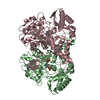
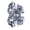
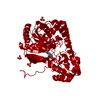
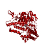
| Title | Crystal structure of the cysteine desulfurase CsdA (persulfurated) from Escherichia coli at 2.384 Angstroem resolution | ||||||
 Components Components | L-CYSTEINE DESULFURASE CSDA | ||||||
 Keywords Keywords | TRANSFERASE / L-CYSTEINE DESULFURASE / SULFUR ACCEPTOR / TRANSPERSULFURATION / SULFUR TRAFFICKING | ||||||
| Function / homology |  Function and homology information Function and homology informationsulfur compound transport / L-selenocysteine catabolic process / Hydrolases; Acting on carbon-sulfur bonds; Acting on carbon-sulfur bonds / cysteine sulfinate desulfinase activity / sulfur amino acid metabolic process / selenocysteine lyase / selenocysteine lyase activity / L-cysteine desulfurase complex / sulfurtransferase activity / L-cysteine catabolic process ...sulfur compound transport / L-selenocysteine catabolic process / Hydrolases; Acting on carbon-sulfur bonds; Acting on carbon-sulfur bonds / cysteine sulfinate desulfinase activity / sulfur amino acid metabolic process / selenocysteine lyase / selenocysteine lyase activity / L-cysteine desulfurase complex / sulfurtransferase activity / L-cysteine catabolic process / cysteine desulfurase / cysteine desulfurase activity / : / iron-sulfur cluster assembly / pyridoxal phosphate binding / hydrolase activity Similarity search - Function | ||||||
| Biological species |  | ||||||
| Method |  X-RAY DIFFRACTION / SYNCHROTRON / MOLECULAR REPLACEMENT / Resolution: 2.384 Å X-RAY DIFFRACTION / SYNCHROTRON / MOLECULAR REPLACEMENT / Resolution: 2.384 Å | ||||||
 Authors Authors | Fernandez, F.J. / Arda, A. / Lopez-Estepa, M. / Aranda, J. / Penya-Soler, E. / Garces, F. / Quintana, J.F. / Round, A. / Campos-Oliva, R. / Bruix, M. ...Fernandez, F.J. / Arda, A. / Lopez-Estepa, M. / Aranda, J. / Penya-Soler, E. / Garces, F. / Quintana, J.F. / Round, A. / Campos-Oliva, R. / Bruix, M. / Coll, M. / Tunon, I. / Jimenez-Barbero, J. / Vega, M.C. | ||||||
 Citation Citation | Journal: Acs Catalysis / Year: 2016 Title: Mechanism of Sulfur Transfer Across Protein-Protein Interfaces: The Cysteine Desulfurase Model System Authors: Fernandez, F.J. / Arda, A. / Lopez-Estepa, M. / Aranda, J. / Penya-Soler, E. / Garces, F. / Quintana, J.F. / Round, A. / Campos-Oliva, R. / Bruix, M. / Coll, M. / Tunon, I. / Jimenez-Barbero, J. / Vega, M.C. | ||||||
| History |
|
- Structure visualization
| Structure viewer | Molecule: MolmilJmol/JSmol |
|---|
- Downloads & links
-Download
| PDBx/mmCIF format | 5ft5.cif.gz | 324 KB | Display | PDBx/mmCIF format |
|---|---|---|---|---|
| PDB format | pdb5ft5.ent.gz | 266 KB | Display | PDB format |
| PDBx/mmJSON format | 5ft5.json.gz | Tree view | PDBx/mmJSON format | |
| Others |  Other downloads Other downloads |
-Validation report
| Arichive directory | https://data.pdbj.org/pub/pdb/validation_reports/ft/5ft5ftp://data.pdbj.org/pub/pdb/validation_reports/ft/5ft5 | HTTPS FTP |
|---|
-Related structure data
| Related structure data |  5ft6C  5ft8C  5ft4S  5ft7 S: Starting model for refinement C: citing same article ( |
|---|---|
| Similar structure data |


-Links
 PDBj
PDBj
- Assembly
| Deposited unit | 
| ||||||||
|---|---|---|---|---|---|---|---|---|---|
| 1 |
| ||||||||
| Unit cell |
| ||||||||
| Noncrystallographic symmetry (NCS) | NCS oper: (Code: given Matrix: (-0.79, -0.4144, -0.4518), Vector: |
-Components
-Protein , 1 types, 2 molecules AB
| #1: Protein | Mass: 43306.047 Da / Num. of mol.: 2 Source method: isolated from a genetically manipulated source Source: (gene. exp.) References: UniProt: Q46925, cysteine desulfurase, Lyases; Carbon-sulfur lyases, selenocysteine lyase |
|---|
-Non-polymers , 5 types, 314 molecules 


| #2: Chemical | ChemComp-GOL /  Mass: 92.094 Da / Num. of mol.: 10 / Source method: obtained synthetically / Formula: C3H8O3 Mass: 92.094 Da / Num. of mol.: 10 / Source method: obtained synthetically / Formula: C3H8O3#3: Chemical | ChemComp-PEG / |  Mass: 106.120 Da / Num. of mol.: 1 / Source method: obtained synthetically / Formula: C4H10O3 Mass: 106.120 Da / Num. of mol.: 1 / Source method: obtained synthetically / Formula: C4H10O3#4: Chemical |  Mass: 150.087 Da / Num. of mol.: 2 / Source method: obtained synthetically / Formula: C4H6O6 Mass: 150.087 Da / Num. of mol.: 2 / Source method: obtained synthetically / Formula: C4H6O6#5: Chemical |  Mass: 247.142 Da / Num. of mol.: 2 / Source method: obtained synthetically / Formula: C8H10NO6P Mass: 247.142 Da / Num. of mol.: 2 / Source method: obtained synthetically / Formula: C8H10NO6P#6: Water | ChemComp-HOH / | Mass: 18.015 Da / Num. of mol.: 299 / Source method: isolated from a natural source / Formula: H2O |
|---|
-Experimental details
-Experiment
| Experiment | Method: X-RAY DIFFRACTION / Number of used crystals: 1 |
|---|
- Sample preparation
| Crystal | Density Matthews: 2.38 Å3/Da / Density % sol: 48.44 % / Description: NONE |
|---|
-Data collection
| Diffraction | Mean temperature: 100 K |
|---|---|
| Diffraction source | Source: SYNCHROTRON / Site: ESRF  / Beamline: ID23-2 / Wavelength: 0.8726 / Beamline: ID23-2 / Wavelength: 0.8726 |
| Detector | Type: ADSC CCD / Detector: CCD |
| Radiation | Protocol: SINGLE WAVELENGTH / Monochromatic (M) / Laue (L): M / Scattering type: x-ray |
| Radiation wavelength | Wavelength: 0.8726 Å / Relative weight: 1 |
| Reflection | Resolution: 2.38→36.72 Å / Num. obs: 29983 / % possible obs: 89.4 % / Observed criterion σ(I): -3 / Redundancy: 4.1 % / Biso Wilson estimate: 22.65 Å2 / Rmerge(I) obs: 0.18 / Net I/σ(I): 11.1 |
| Reflection shell | Resolution: 2.38→2.47 Å / Redundancy: 4.1 % / Rmerge(I) obs: 0.74 / Mean I/σ(I) obs: 2.4 / % possible all: 91.5 |
- Processing
| Software |
| ||||||||||||||||||||||||||||||||||||||||||||||||||||||||||||||||||||||||||||||||||||
|---|---|---|---|---|---|---|---|---|---|---|---|---|---|---|---|---|---|---|---|---|---|---|---|---|---|---|---|---|---|---|---|---|---|---|---|---|---|---|---|---|---|---|---|---|---|---|---|---|---|---|---|---|---|---|---|---|---|---|---|---|---|---|---|---|---|---|---|---|---|---|---|---|---|---|---|---|---|---|---|---|---|---|---|---|---|
| Refinement | Method to determine structure: MOLECULAR REPLACEMENT Starting model: PDB ENTRY 5FT4 Resolution: 2.384→36.721 Å / SU ML: 0.27 / σ(F): 1.9 / Phase error: 21.21 / Stereochemistry target values: ML / Details: U VALUES WITH TLS ADDED.
| ||||||||||||||||||||||||||||||||||||||||||||||||||||||||||||||||||||||||||||||||||||
| Solvent computation | Shrinkage radii: 0.9 Å / VDW probe radii: 1.11 Å / Solvent model: FLAT BULK SOLVENT MODEL | ||||||||||||||||||||||||||||||||||||||||||||||||||||||||||||||||||||||||||||||||||||
| Displacement parameters | Biso mean: 30.5 Å2 | ||||||||||||||||||||||||||||||||||||||||||||||||||||||||||||||||||||||||||||||||||||
| Refinement step | Cycle: LAST / Resolution: 2.384→36.721 Å
| ||||||||||||||||||||||||||||||||||||||||||||||||||||||||||||||||||||||||||||||||||||
| Refine LS restraints |
| ||||||||||||||||||||||||||||||||||||||||||||||||||||||||||||||||||||||||||||||||||||
| LS refinement shell |
| ||||||||||||||||||||||||||||||||||||||||||||||||||||||||||||||||||||||||||||||||||||
| Refinement TLS params. | Method: refined / Origin x: 21.4065 Å / Origin y: 44.2324 Å / Origin z: 17.57 Å
| ||||||||||||||||||||||||||||||||||||||||||||||||||||||||||||||||||||||||||||||||||||
| Refinement TLS group | Selection details: ALL |
