[English] 日本語
Yorodumi
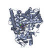
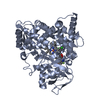
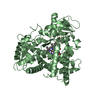
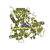
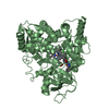
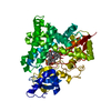
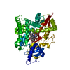
- PDB-5frb: Crystal structure of sterol 14-alpha demethylase (CYP51B) from a ... -

+
Open data


ID or keywords:

Loading...

-
Basic information

Entry
Database: PDB / ID: 5frb
TitleCrystal structure of sterol 14-alpha demethylase (CYP51B) from a pathogenic filamentous fungus Aspergillus fumigatus in complex with a tetrazole-based inhibitor VT-1598
ComponentsSTEROL 14-ALPHA DEMETHYLASE, CYP51B
KeywordsOXIDOREDUCTASE / STEROL 14-ALPHA DEMETHYLASE (CYP51) / CYTOCHROME P450 / HEME / MONOOXYGENASE / STEROL BIOSYNTHESIS / EUKARYOTIC MEMBRANES / CYTOCHROME P450 FOLD / ENDOPLASMIC RETICULUM MEMBRANE / OXIDOREDUCTASE-OXIDOREDUCTASE INHIBITOR COMPLEX
Function / homology
Function and homology information


sterol 14alpha-demethylase / sterol 14-demethylase activity / ergosterol biosynthetic process / oxidoreductase activity, acting on paired donors, with incorporation or reduction of molecular oxygen / monooxygenase activity / methyltransferase activity / membrane => GO:0016020 / methylation / oxidoreductase activity / iron ion binding ...sterol 14alpha-demethylase / sterol 14-demethylase activity / ergosterol biosynthetic process / oxidoreductase activity, acting on paired donors, with incorporation or reduction of molecular oxygen / monooxygenase activity / methyltransferase activity / membrane => GO:0016020 / methylation / oxidoreductase activity / iron ion binding / heme binding / endoplasmic reticulum membrane
Similarity search - Function
: / Cytochrome P450, E-class, group IV / Cytochrome P450, conserved site / Cytochrome P450 cysteine heme-iron ligand signature. / Cytochrome P450 / Cytochrome P450 superfamily / Cytochrome P450
Similarity search - Domain/homology
PROTOPORPHYRIN IX CONTAINING FE / Chem-VT2 / 14-alpha sterol demethylase Cyp51B / Sterol 14-alpha demethylase cyp51B
Similarity search - Component
Biological speciesASPERGILLUS FUMIGATUS (mold)
MethodX-RAY DIFFRACTION / SYNCHROTRON / MOLECULAR REPLACEMENT / Resolution: 2.99 Å
AuthorsHargrove, T.Y. / Wawrzak, Z. / Lepesheva, G.I.
CitationJournal: Antimicrob. Agents Chemother. / Year: 2017
Title: Crystal Structure of the New Investigational Drug Candidate VT-1598 in Complex with Aspergillus fumigatus Sterol 14 alpha-Demethylase Provides Insights into Its Broad-Spectrum Antifungal Activity.
Authors: Hargrove, T.Y. / Garvey, E.P. / Hoekstra, W.J. / Yates, C.M. / Wawrzak, Z. / Rachakonda, G. / Villalta, F. / Lepesheva, G.I.
History
DepositionDec 16, 2015Deposition site: PDBE / Processing site: PDBE
Revision 1.0Apr 26, 2017Provider: repository / Type: Initial release
Revision 1.1May 10, 2017Group: Database references
Revision 1.2May 17, 2017Group: Database references
Revision 1.3Jul 5, 2017Group: Database references / Category: citation
Item: _citation.country / _citation.journal_id_ASTM ..._citation.country / _citation.journal_id_ASTM / _citation.journal_id_CSD / _citation.journal_volume / _citation.title
Revision 1.4Jan 10, 2024Group: Data collection / Database references ...Data collection / Database references / Derived calculations / Other / Refinement description
Category: chem_comp_atom / chem_comp_bond ...chem_comp_atom / chem_comp_bond / database_2 / pdbx_database_status / pdbx_initial_refinement_model / struct_site
Item: _database_2.pdbx_DOI / _database_2.pdbx_database_accession ..._database_2.pdbx_DOI / _database_2.pdbx_database_accession / _pdbx_database_status.status_code_sf / _struct_site.pdbx_auth_asym_id / _struct_site.pdbx_auth_comp_id / _struct_site.pdbx_auth_seq_id

-
Structure visualization

Structure viewerMolecule:
MolmilJmol/JSmol

Downloads & links

-
Assembly

Deposited unit
A: STEROL 14-ALPHA DEMETHYLASE, CYP51B
hetero molecules


Theoretical massNumber of molelcules
Total (without water)54,7843
Polymers53,5811
Non-polymers1,2032
Water00
1


  • Idetical with deposited unit
  • defined by author&software
TypeNameSymmetry operationNumber
identity operation1_555x,y,z1
MethodPISA
Unit cell
Length a, b, c (Å)109.857, 109.857, 84.838
Angle α, β, γ (deg.)90.00, 90.00, 120.00
Int Tables number152
Space group name H-MP3121

-
Components

#1: Protein STEROL 14-ALPHA DEMETHYLASE, CYP51B


Mass: 53581.266 Da / Num. of mol.: 1 / Fragment: CATALYTIC DOMAIN, RESIDUES 50-519 / Mutation: YES
Source method: isolated from a genetically manipulated source
Source: (gene. exp.) ASPERGILLUS FUMIGATUS (mold) / Gene: ERG 11 / Plasmid: PCW / Production host: ESCHERICHIA COLI (E. coli) / Strain (production host): HMS174
References: UniProt: A0A0J5PJX1, UniProt: E9QY26*PLUS, sterol 14alpha-demethylase
#2: Chemical ChemComp-HEM / PROTOPORPHYRIN IX CONTAINING FE / HEME


Mass: 616.487 Da / Num. of mol.: 1 / Source method: obtained synthetically / Formula: C34H32FeN4O4
#3: Chemical ChemComp-VT2 / (R)-4-((4-((6-(2-(2,4-difluorophenyl)-1,1-difluoro-2-hydroxy-3-(1H-tetrazol-1-yl)propyl)pyridin-3-yl)ethynyl)phenoxy)methyl)benzonitrile


Mass: 586.539 Da / Num. of mol.: 1 / Source method: obtained synthetically / Formula: C31H22F4N6O2

-
Experimental details

-
Experiment

ExperimentMethod: X-RAY DIFFRACTION / Number of used crystals: 1

-
Sample preparation

CrystalDensity Matthews: 2.77 Å3/Da / Density % sol: 55.6 % / Description: NONE
Crystal growpH: 7.4 / Details: pH 7.4

-
Data collection

DiffractionMean temperature: 100 K
Diffraction sourceSource: SYNCHROTRON / Site: APS / Beamline: 21-ID-F / Wavelength: 0.97856
DetectorType: MARRESEARCH / Detector: CCD / Date: Sep 25, 2015 / Details: BE LENSES DIAMOND LAUE
RadiationMonochromator: DIAMOND / Protocol: SINGLE WAVELENGTH / Monochromatic (M) / Laue (L): M / Scattering type: x-ray
Radiation wavelengthWavelength: 0.97856 Å / Relative weight: 1
ReflectionResolution: 2.99→95.15 Å / Num. obs: 12238 / % possible obs: 99.7 % / Observed criterion σ(I): 2 / Redundancy: 5.4 % / Biso Wilson estimate: 116.3 Å2 / Rmerge(I) obs: 0.03 / Net I/σ(I): 26.1
Reflection shellResolution: 2.99→3.07 Å / Redundancy: 5.6 % / Rmerge(I) obs: 0.65 / Mean I/σ(I) obs: 1.9 / % possible all: 100

-
Processing

Software
NameVersionClassification
REFMAC5.8.0131refinement
MOSFLMdata reduction
SCALEPACKdata scaling
PHASERphasing
RefinementMethod to determine structure: MOLECULAR REPLACEMENT
Starting model: PDB ENTRY 4UHL
Resolution: 2.99→95.14 Å / Cor.coef. Fo:Fc: 0.942 / Cor.coef. Fo:Fc free: 0.936 / SU B: 67.354 / SU ML: 0.523 / Cross valid method: THROUGHOUT / ESU R Free: 0.496 / Stereochemistry target values: MAXIMUM LIKELIHOOD
Details: HYDROGENS HAVE BEEN ADDED IN THE RIDING POSITIONS. U VALUES WITH TLS ADDED
RfactorNum. reflection% reflectionSelection details
Rfree0.28006 674 5.6 %RANDOM
Rwork0.25839 ---
obs0.25955 11467 99.22 %-
Solvent computationIon probe radii: 0.8 Å / Shrinkage radii: 0.8 Å / VDW probe radii: 1.2 Å / Solvent model: MASK
Displacement parametersBiso mean: 153.732 Å2
Baniso -1Baniso -2Baniso -3
1-0.19 Å20.09 Å20 Å2
2--0.19 Å2-0 Å2
3----0.61 Å2
Refinement stepCycle: LAST / Resolution: 2.99→95.14 Å
ProteinNucleic acidLigandSolventTotal
Num. atoms3752 0 86 0 3838
Refine LS restraints
Refine-IDTypeDev idealDev ideal targetNumber
X-RAY DIFFRACTIONr_bond_refined_d0.0050.023951
X-RAY DIFFRACTIONr_bond_other_d00.023700
X-RAY DIFFRACTIONr_angle_refined_deg0.7541.9885378
X-RAY DIFFRACTIONr_angle_other_deg0.5263.0028523
X-RAY DIFFRACTIONr_dihedral_angle_1_deg5.7995469
X-RAY DIFFRACTIONr_dihedral_angle_2_deg30.39423.237173
X-RAY DIFFRACTIONr_dihedral_angle_3_deg14.80215648
X-RAY DIFFRACTIONr_dihedral_angle_4_deg14.141525
X-RAY DIFFRACTIONr_chiral_restr0.0470.2573
X-RAY DIFFRACTIONr_gen_planes_refined0.0070.0214418
X-RAY DIFFRACTIONr_gen_planes_other0.0020.02936
X-RAY DIFFRACTIONr_nbd_refined
X-RAY DIFFRACTIONr_nbd_other
X-RAY DIFFRACTIONr_nbtor_refined
X-RAY DIFFRACTIONr_nbtor_other
X-RAY DIFFRACTIONr_xyhbond_nbd_refined
X-RAY DIFFRACTIONr_xyhbond_nbd_other
X-RAY DIFFRACTIONr_metal_ion_refined
X-RAY DIFFRACTIONr_metal_ion_other
X-RAY DIFFRACTIONr_symmetry_vdw_refined
X-RAY DIFFRACTIONr_symmetry_vdw_other
X-RAY DIFFRACTIONr_symmetry_hbond_refined
X-RAY DIFFRACTIONr_symmetry_hbond_other
X-RAY DIFFRACTIONr_symmetry_metal_ion_refined
X-RAY DIFFRACTIONr_symmetry_metal_ion_other
X-RAY DIFFRACTIONr_mcbond_it14.54411.5971879
X-RAY DIFFRACTIONr_mcbond_other14.54711.5931878
X-RAY DIFFRACTIONr_mcangle_it20.41417.4572347
X-RAY DIFFRACTIONr_mcangle_other
X-RAY DIFFRACTIONr_scbond_it14.30212.2562072
X-RAY DIFFRACTIONr_scbond_other
X-RAY DIFFRACTIONr_scangle_it
X-RAY DIFFRACTIONr_scangle_other
X-RAY DIFFRACTIONr_long_range_B_refined
X-RAY DIFFRACTIONr_long_range_B_other
X-RAY DIFFRACTIONr_rigid_bond_restr
X-RAY DIFFRACTIONr_sphericity_free
X-RAY DIFFRACTIONr_sphericity_bonded
LS refinement shellResolution: 2.995→3.072 Å / Total num. of bins used: 20
RfactorNum. reflection% reflection
Rfree0.359 62 -
Rwork0.339 813 -
obs--97.77 %
Refinement TLS params.Method: refined / Origin x: 293.3559 Å / Origin y: 16.6827 Å / Origin z: 8.3265 Å
111213212223313233
T0.4704 Å20.1908 Å2-0.1167 Å2-0.1971 Å2-0.1747 Å2--0.3436 Å2
L1.1223 °2-0.3911 °20.0508 °2-1.8984 °2-0.5923 °2--0.2513 °2
S0.5967 Å °0.1397 Å °-0.0702 Å °-0.2405 Å °-0.311 Å °0.7588 Å °0.0858 Å °0.1768 Å °-0.2858 Å °

+
About Yorodumi

-
News

-
Feb 9, 2022. New format data for meta-information of EMDB entries

New format data for meta-information of EMDB entries

  • Version 3 of the EMDB header file is now the official format.
  • The previous official version 1.9 will be removed from the archive.

Related info.:EMDB header

External links:wwPDB to switch to version 3 of the EMDB data model

-
Aug 12, 2020. Covid-19 info

Covid-19 info

URL: https://pdbj.org/emnavi/covid19.php

New page: Covid-19 featured information page in EM Navigator.

Related info.:Covid-19 info / Mar 5, 2020. Novel coronavirus structure data

+
Mar 5, 2020. Novel coronavirus structure data

Novel coronavirus structure data

Related info.:Yorodumi Speices / Aug 12, 2020. Covid-19 info

External links:COVID-19 featured content - PDBj / Molecule of the Month (242):Coronavirus Proteases

+
Jan 31, 2019. EMDB accession codes are about to change! (news from PDBe EMDB page)

EMDB accession codes are about to change! (news from PDBe EMDB page)

  • The allocation of 4 digits for EMDB accession codes will soon come to an end. Whilst these codes will remain in use, new EMDB accession codes will include an additional digit and will expand incrementally as the available range of codes is exhausted. The current 4-digit format prefixed with “EMD-” (i.e. EMD-XXXX) will advance to a 5-digit format (i.e. EMD-XXXXX), and so on. It is currently estimated that the 4-digit codes will be depleted around Spring 2019, at which point the 5-digit format will come into force.
  • The EM Navigator/Yorodumi systems omit the EMD- prefix.

Related info.:Q: What is EMD? / ID/Accession-code notation in Yorodumi/EM Navigator

External links:EMDB Accession Codes are Changing Soon! / Contact to PDBj

+
Jul 12, 2017. Major update of PDB

Major update of PDB

  • wwPDB released updated PDB data conforming to the new PDBx/mmCIF dictionary.
  • This is a major update changing the version number from 4 to 5, and with Remediation, in which all the entries are updated.
  • In this update, many items about electron microscopy experimental information are reorganized (e.g. em_software).
  • Now, EM Navigator and Yorodumi are based on the updated data.

External links:wwPDB Remediation / Enriched Model Files Conforming to OneDep Data Standards Now Available in the PDB FTP Archive

-
Yorodumi

Thousand views of thousand structures

  • Yorodumi is a browser for structure data from EMDB, PDB, SASBDB, etc.
  • This page is also the successor to EM Navigator detail page, and also detail information page/front-end page for Omokage search.
  • The word "yorodu" (or yorozu) is an old Japanese word meaning "ten thousand". "mi" (miru) is to see.

Related info.:EMDB / PDB / SASBDB / Comparison of 3 databanks / Yorodumi Search / Aug 31, 2016. New EM Navigator & Yorodumi / Yorodumi Papers / Jmol/JSmol / Function and homology information / Changes in new EM Navigator and Yorodumi

Read more