 Movie
Movie Controller

[English] 日本語
 Yorodumi
- PDB-5fq8: Crystal structure of the SusCD complex BT2261-2264 from Bacteroid... -
+ Open data

- Basic information
| Entry | Database: PDB / ID: 5fq8 | ||||||
|---|---|---|---|---|---|---|---|
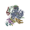
| Title | Crystal structure of the SusCD complex BT2261-2264 from Bacteroides thetaiotaomicron | ||||||
 Components Components |
| ||||||
 Keywords Keywords | MEMBRANE PROTEIN / OUTER MEMBRANE NUTRIENT IMPORTER SUSCD COMPLEX | ||||||
| Function / homology |  Function and homology information Function and homology information | ||||||
| Biological species |  BACTEROIDES THETAIOTAOMICRON (bacteria) BACTEROIDES THETAIOTAOMICRON (bacteria) | ||||||
| Method |  X-RAY DIFFRACTION / SYNCHROTRON / MOLECULAR REPLACEMENT / Resolution: 2.75 Å X-RAY DIFFRACTION / SYNCHROTRON / MOLECULAR REPLACEMENT / Resolution: 2.75 Å | ||||||
 Authors Authors | Glenwright, A.J. / Pothula, K.R. / Chorev, D.S. / Basle, A. / Robinson, C.V. / Kleinekathoefer, U. / Bolam, D.N. / van den Berg, B. | ||||||
 Citation Citation | Journal: Nature / Year: 2017 Title: Structural basis for nutrient acquisition by dominant members of the human gut microbiota. Authors: Glenwright, A.J. / Pothula, K.R. / Bhamidimarri, S.P. / Chorev, D.S. / Basle, A. / Firbank, S.J. / Zheng, H. / Robinson, C.V. / Winterhalter, M. / Kleinekathofer, U. / Bolam, D.N. / van den Berg, B. | ||||||
| History |
| ||||||
| Remark 700 | SHEET DETERMINATION METHOD: DSSP THE SHEETS PRESENTED AS "BD" IN EACH CHAIN ON SHEET RECORDS BELOW ... SHEET DETERMINATION METHOD: DSSP THE SHEETS PRESENTED AS "BD" IN EACH CHAIN ON SHEET RECORDS BELOW IS ACTUALLY AN 22-STRANDED BARREL THIS IS REPRESENTED BY A 23-STRANDED SHEET IN WHICH THE FIRST AND LAST STRANDS ARE IDENTICAL. SHEET DETERMINATION METHOD: DSSP THE SHEETS PRESENTED AS "DD" IN EACH CHAIN ON SHEET RECORDS BELOW IS ACTUALLY AN 22-STRANDED BARREL THIS IS REPRESENTED BY A 23-STRANDED SHEET IN WHICH THE FIRST AND LAST STRANDS ARE IDENTICAL. |
- Structure visualization
| Structure viewer | Molecule: MolmilJmol/JSmol |
|---|
- Downloads & links
-Download
| PDBx/mmCIF format | 5fq8.cif.gz | 1.3 MB | Display | PDBx/mmCIF format |
|---|---|---|---|---|
| PDB format | pdb5fq8.ent.gz | 1.1 MB | Display | PDB format |
| PDBx/mmJSON format | 5fq8.json.gz | Tree view | PDBx/mmJSON format | |
| Others |  Other downloads Other downloads |
-Validation report
| Arichive directory | https://data.pdbj.org/pub/pdb/validation_reports/fq/5fq8ftp://data.pdbj.org/pub/pdb/validation_reports/fq/5fq8 | HTTPS FTP |
|---|
-Related structure data
| Related structure data |  5fq3C  5fq4SC  5fq6C  5fq7C  5lx8C  5t3rC  5t4yC  1fepS  3h3iS C: citing same article ( S: Starting model for refinement |
|---|---|
| Similar structure data |

-Links
 PDBj
PDBj


- Assembly
| Deposited unit | 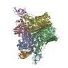
| ||||||||
|---|---|---|---|---|---|---|---|---|---|
| 1 |
| ||||||||
| Unit cell |
|
-Components
-Protein , 4 types, 7 molecules ACBDEFG
| #1: Protein | Mass: 53145.875 Da / Num. of mol.: 2 / Source method: isolated from a natural source / Details: OUTER MEMBRANE LIPOPROTEIN / Source: (natural) BACTEROIDES THETAIOTAOMICRON (bacteria) / References: UniProt: Q8A5H6#2: Protein | Mass: 108954.195 Da / Num. of mol.: 2 / Source method: isolated from a natural source / Details: TONB DEPENDENT TRANSPORTER / Source: (natural) BACTEROIDES THETAIOTAOMICRON (bacteria) / References: UniProt: Q8A5H5#3: Protein | Mass: 16428.965 Da / Num. of mol.: 2 / Source method: isolated from a natural source / Details: OUTER MEMBRANE LIPOPROTEIN / Source: (natural) BACTEROIDES THETAIOTAOMICRON (bacteria) / References: UniProt: Q8A5H8#4: Protein | | Mass: 23399.908 Da / Num. of mol.: 1 / Source method: isolated from a natural source / Details: OUTER MEMBRANE LIPOPROTEIN / Source: (natural) BACTEROIDES THETAIOTAOMICRON (bacteria) / References: UniProt: Q8A5H7 |
|---|
-UNCHARACTERISED PROTEIN, BOUND ... , 2 types, 2 molecules PQ
| #5: Protein/peptide | Mass: 588.533 Da / Num. of mol.: 1 / Source method: isolated from a natural source / Details: OUTER MEMBRANE LIPOPROTEIN / Source: (natural) BACTEROIDES THETAIOTAOMICRON (bacteria) |
|---|---|
| #6: Protein/peptide | Mass: 531.481 Da / Num. of mol.: 1 / Source method: isolated from a natural source / Details: OUTER MEMBRANE LIPOPROTEIN / Source: (natural) BACTEROIDES THETAIOTAOMICRON (bacteria) |
-Non-polymers , 5 types, 942 molecules 


| #7: Chemical | ChemComp-MG /  Mass: 24.305 Da / Num. of mol.: 4 / Source method: obtained synthetically / Formula: Mg Mass: 24.305 Da / Num. of mol.: 4 / Source method: obtained synthetically / Formula: Mg#8: Chemical |  Mass: 384.593 Da / Num. of mol.: 2 / Source method: obtained synthetically / Formula: C23H44O4 Mass: 384.593 Da / Num. of mol.: 2 / Source method: obtained synthetically / Formula: C23H44O4#9: Chemical | ChemComp-CA /  Mass: 40.078 Da / Num. of mol.: 4 / Source method: obtained synthetically / Formula: Ca Mass: 40.078 Da / Num. of mol.: 4 / Source method: obtained synthetically / Formula: Ca#10: Chemical | ChemComp-C8E / (  Mass: 306.438 Da / Num. of mol.: 4 / Source method: obtained synthetically / Formula: C16H34O5 / Comment: C8E, detergent*YM Mass: 306.438 Da / Num. of mol.: 4 / Source method: obtained synthetically / Formula: C16H34O5 / Comment: C8E, detergent*YM#11: Water | ChemComp-HOH / | Mass: 18.015 Da / Num. of mol.: 928 / Source method: isolated from a natural source / Formula: H2O |
|---|
-Details
| Has protein modification | Y |
|---|---|
| Sequence details | IDENTITY OF PEPTIDE LIGAND UNKNOWN. ASSIGNED SEQUENCE FITS REASONABLY WELL IN DENSITY. LIKELY AN ...IDENTITY OF PEPTIDE LIGAND UNKNOWN. ASSIGNED SEQUENCE FITS REASONABLY |
-Experimental details
-Experiment
| Experiment | Method: X-RAY DIFFRACTION / Number of used crystals: 1 |
|---|
- Sample preparation
| Crystal | Density Matthews: 3.1 Å3/Da / Density % sol: 60 % / Description: NONE |
|---|---|
| Crystal grow | Details: 0.2 M MAGNESIUM FORMATE DIHYDRATE 0.05 M TRIS PH 8 18-22% PEG3350 |
-Data collection
| Diffraction | Mean temperature: 100 K |
|---|---|
| Diffraction source | Source: SYNCHROTRON / Site: Diamond  / Beamline: I04 / Wavelength: 0.9795 / Beamline: I04 / Wavelength: 0.9795 |
| Detector | Type: DECTRIS PIXEL / Detector: PIXEL / Date: Jan 31, 2015 |
| Radiation | Protocol: SINGLE WAVELENGTH / Monochromatic (M) / Laue (L): M / Scattering type: x-ray |
| Radiation wavelength | Wavelength: 0.9795 Å / Relative weight: 1 |
| Reflection | Resolution: 2.75→66.95 Å / Num. obs: 125711 / % possible obs: 100 % / Observed criterion σ(I): -3 / Redundancy: 3.8 % / Rmerge(I) obs: 0.19 / Net I/σ(I): 5.8 |
| Reflection shell | Resolution: 2.75→2.8 Å / Redundancy: 3.6 % / Rmerge(I) obs: 0.66 / Mean I/σ(I) obs: 1.7 / % possible all: 100 |
- Processing
| Software |
| ||||||||||||||||||||||||||||||||||||||||||||||||||||||||||||||||||||||||||||||||||||||||||||||||||||||||||||||||||||||||||||||||||||||||||||||||||||||||||||||||||||||||||||||||||||||||||||||||||||||||||||||||||||||||||||||||||||||||||||||||||||||||||
|---|---|---|---|---|---|---|---|---|---|---|---|---|---|---|---|---|---|---|---|---|---|---|---|---|---|---|---|---|---|---|---|---|---|---|---|---|---|---|---|---|---|---|---|---|---|---|---|---|---|---|---|---|---|---|---|---|---|---|---|---|---|---|---|---|---|---|---|---|---|---|---|---|---|---|---|---|---|---|---|---|---|---|---|---|---|---|---|---|---|---|---|---|---|---|---|---|---|---|---|---|---|---|---|---|---|---|---|---|---|---|---|---|---|---|---|---|---|---|---|---|---|---|---|---|---|---|---|---|---|---|---|---|---|---|---|---|---|---|---|---|---|---|---|---|---|---|---|---|---|---|---|---|---|---|---|---|---|---|---|---|---|---|---|---|---|---|---|---|---|---|---|---|---|---|---|---|---|---|---|---|---|---|---|---|---|---|---|---|---|---|---|---|---|---|---|---|---|---|---|---|---|---|---|---|---|---|---|---|---|---|---|---|---|---|---|---|---|---|---|---|---|---|---|---|---|---|---|---|---|---|---|---|---|---|---|---|---|---|---|---|---|---|---|---|---|---|---|---|---|---|---|
| Refinement | Method to determine structure: MOLECULAR REPLACEMENT Starting model: PDB ENTRIES 5FQ4, 3H3I, 1FEP Resolution: 2.75→52.933 Å / SU ML: 0.39 / σ(F): 1.96 / Phase error: 27.39 / Stereochemistry target values: ML
| ||||||||||||||||||||||||||||||||||||||||||||||||||||||||||||||||||||||||||||||||||||||||||||||||||||||||||||||||||||||||||||||||||||||||||||||||||||||||||||||||||||||||||||||||||||||||||||||||||||||||||||||||||||||||||||||||||||||||||||||||||||||||||
| Solvent computation | Shrinkage radii: 0.9 Å / VDW probe radii: 1.11 Å / Solvent model: FLAT BULK SOLVENT MODEL | ||||||||||||||||||||||||||||||||||||||||||||||||||||||||||||||||||||||||||||||||||||||||||||||||||||||||||||||||||||||||||||||||||||||||||||||||||||||||||||||||||||||||||||||||||||||||||||||||||||||||||||||||||||||||||||||||||||||||||||||||||||||||||
| Refinement step | Cycle: LAST / Resolution: 2.75→52.933 Å
| ||||||||||||||||||||||||||||||||||||||||||||||||||||||||||||||||||||||||||||||||||||||||||||||||||||||||||||||||||||||||||||||||||||||||||||||||||||||||||||||||||||||||||||||||||||||||||||||||||||||||||||||||||||||||||||||||||||||||||||||||||||||||||
| Refine LS restraints |
| ||||||||||||||||||||||||||||||||||||||||||||||||||||||||||||||||||||||||||||||||||||||||||||||||||||||||||||||||||||||||||||||||||||||||||||||||||||||||||||||||||||||||||||||||||||||||||||||||||||||||||||||||||||||||||||||||||||||||||||||||||||||||||
| LS refinement shell |
| ||||||||||||||||||||||||||||||||||||||||||||||||||||||||||||||||||||||||||||||||||||||||||||||||||||||||||||||||||||||||||||||||||||||||||||||||||||||||||||||||||||||||||||||||||||||||||||||||||||||||||||||||||||||||||||||||||||||||||||||||||||||||||
| Refinement TLS params. | Method: refined / Refine-ID: X-RAY DIFFRACTION
| ||||||||||||||||||||||||||||||||||||||||||||||||||||||||||||||||||||||||||||||||||||||||||||||||||||||||||||||||||||||||||||||||||||||||||||||||||||||||||||||||||||||||||||||||||||||||||||||||||||||||||||||||||||||||||||||||||||||||||||||||||||||||||
| Refinement TLS group |
|
